Article Text

Statistics from Altmetric.com
- electromyogram
- nerve conduction studies
- nerve action potential
- Lambert Eaton myasthenic syndrome
- neuromuscular transmission disorders
A neurologist has sent a patient for nerve conduction studies (NCS) and has received the report, but what does it mean? We hope to remove some of the mysteries that may surround NCS. The techniques and how they are affected by disease are described in general terms. These principles can be applied to specific conditions discussed elsewhere. We also discuss the numerous pitfalls that may be encountered with NCS. Understanding these basic concepts will allow you to get the most from your clinical neurophysiology department.
NCS are only part of a complete peripheral neurophysiological examination (PNE) and are frequently accompanied by a needle electromyogram (EMG). The combination of both techniques and those detailed in other articles in this issue are often required for a complete diagnostic study. The process of choosing the appropriate tests is the responsibility of the clinical neurophysiologist (CN) and not the referring doctor and is planned as a dynamic series of steps designed to answer specific questions about nervous system function raised by the clinical picture.
ABBREVIATIONS
Clinical neurophysiologists can employ a confusing number of terms and abbreviations. Box 1 lists the ones we use frequently.
Box 1: List of abbreviations and terminology used
-
ACh: acetylcholine
-
AIDP: acute inflammatory demyelinating polyneuropathy
-
AMAN: acute motor axonal neuropathy
-
CMAP: compound muscle action potential
-
CN: clinical neurophysiologist
-
DRG: dorsal root ganglion
-
EMG: electromyogram
-
LEMS: Lambert-Eaton myasthenic syndrome
-
MG: myasthenia gravis
-
NAP: nerve action potential
-
NCS: nerve conduction studies
-
NMTD: neuromuscular transmission disorders
-
PNE: peripheral neurophysiological examination
-
PNS: peripheral nervous system
-
RNS: repetitive nerve stimulation
-
SNAP: sensory nerve action potential
-
TMS: transcranial magnetic stimulation
-
Conduction block: A reduction of proximal CMAP area/amplitude of at least 20% (usually > 50%) compared with distal CMAP area/amplitude. The duration of the proximal CMAP should not increase by > 20% (see temporal dispersion)
-
Temporal dispersion: A reduction in proximal CMAP amplitude compared with distal CMAP amplitude when the proximal CMAP duration increases by > 20%
-
Orthodromic: Nerve action potentials carried in the physiological direction
-
Antidromic: Nerve action potentials carried in the direction opposite to the physiological
WHAT DO YOU TELL THE PATIENT ABOUT THE TESTS?
NCS involve activating nerves electrically with small safe pulses over several points on the skin of the limbs and measuring the responses obtained. Some people are frightened by the term “electric shocks”. The tests are at worst uncomfortable but most patients find them tolerable when they are explained sympathetically. There are no long term side effects. Both authors have had their own nerves tested on many occasions without difficulty and neither of us would claim particularly high pain thresholds. A self inflicted demonstration by the CN may be helpful with the more anxious patient as well as a simple information sheet.
There are very few contraindications to these investigations, but the most important is the presence of some cardiac pacemakers. With most there is no risk but discussion with the patient’s cardiologist is advised if (1) the NCS are likely to involve stimulation close to the chest wall, and (2) if a life threatening event would be risked should the pacemaker either be triggered or changed to a harmful default state if subject to an external voltage.
REFERRING PATIENTS FOR NERVE CONDUCTION STUDIES
NCS give data on peripheral nervous system (PNS) function which may be used to provide:
-
diagnosis(es)
-
description of disease state (old/new; dynamic/static pathophysiology)
-
longitudinal monitoring of disease with multiple studies
-
advice on prognosis and management based on tests results and disease detected.
Nerve conduction studies may be diagnostically helpful in patients suspected of having almost any PNS disorder including disorders of nerve roots, peripheral nerves, muscle and neuromuscular junction. Cranial nerves and spinal cord function may also be assessed. Specific clinical indications are discussed elsewhere.
When referring patients, it is useful to think of the specific questions you wish answered with the PNE. In difficult cases it is helpful to discuss the referral (which should contain all relevant clinical information) with the CN. The PNE is an extension of the clinical history and examination and CNs will take a history and perform the relevant neurological examination but will rely on the referral information to guide them. In straightforward conditions they may follow an initial standard protocol of tests but the investigator will be ready to modify or add to these tests on the basis of the initial findings. This emphasises that when NCS are performed by technical staff, the CN should supervise in close proximity and be available to carry out other appropriate tests. It is unnecessary (and sometimes insulting) to specify tests in the referral as long as the clinical question being asked is clear. For example, in a patient suspected of having right carpal tunnel syndrome it is unnecessary to ask for “NCS of the right median nerve”. (The wise CN will study both median nerves anyway as this condition is frequently bilateral.)
Basic nerve and muscle physiology
The referring doctor needs only a minimal knowledge of basic and applied physiology to understand the test results, but all PNE reports should be written in terms that do not assume specialist knowledge. A minimum knowledge set to understand the principles of the techniques is shown in box 2 with links to more detail.
Box 2: Minimum basic physiological knowledge to understand nerve conduction studies
-
Membrane potential: genesis; threshold effects; effect of membrane damage; denervation
-
Single axon: saltatory and non-saltatory conduction; factors which determine conduction velocity: diameter, myelination, inter-nodal distance
-
Whole nerve composition: fascicular structure; size and conduction velocity distribution; afferent and efferent modalities and their relative contribution
-
Neuromuscular transmission: nerve terminal function; transmitter production, storage and release; post-synaptic membrane structure and function; receptor dynamics; endplate potentials; propagated muscle action potentials
-
External electrical/magnetic stimulation: local depolarisation; bidirectional propagation once depolarised; effects of skin and subcutaneous tissue impedance; electrical inductive effect of applied magnetic field; stimulation threshold depends on: membrane properties(accommodation), nerve fibre location and size with respect to stimulator
-
Muscle function: excitation contraction coupling; muscle fibre types and function; fatigue
For an excellent interactive physiological education tool see Richard Carpenter’s Neurolab at http://www.cudos.ac.uk/web/copyright.htm.
The principals of nerve conduction studies
NCS involve the application of a depolarising square wave electrical pulses to the skin over a peripheral nerve producing: (1) a propagated nerve action potential (NAP) recorded at a distant point over the same nerve: and (2) a compound muscle action potential (CMAP) arising from the activation of muscle fibres in a target muscle supplied by the nerve. In both cases these may be recorded with surface or needle electrodes.
Surface electrodes are designed to give information about the whole of a muscle stimulated, giving data for the time taken for the fastest axons to conduct an impulse to the muscle and the size of the response.
Needle electrodes for NCS give very accurate conduction time information, but because they record from only a small area of muscle or nerve, they give poor or, in the case of the latter, more complex information making numerical analysis difficult. However, needle recordings are most appropriate when severe muscle wasting has occurred, or when the depth of a muscle under study makes a surface recording impossible.
Nerves may be stimulated through the skin with surface stimulators, or via a needle placed close to a nerve or a nerve root. Spinal root and cerebral cortical stimulation may also be carried out using transcutaneous magnetic stimulation (TMS) dealt with elsewhere in this issue. Thus the full length of the motor pathway may be assessed from cortex to cord, root, neuromuscular junction, and the contractile apparatus. Choice of the stimulation points depends both on the desire to “bracket” above and below the point of a proposed focal lesion and the anatomical availability of the appropriate structure.
The aim of the NCS
Our minimum knowledge set above has shown us that peripheral nerves contain many nerve fibres of different diameters, degrees of myelination, and afferent or efferent connections. The NCS studies the fastest 20% of these fibres and the aim of the investigation is to document focal or continuous abnormalities in the length of the mixed, motor or sensory nerve. Particular attention is paid to the following questions as the test progresses:
-
Is the fastest conduction velocity normal?
-
Is the velocity gradient normal. Normally nerves closer to the neuraxis and more cephalad conduct faster than more distal and caudal nerves.
-
Is the CMAP normal in size and shape?
-
Does the CMAP alter in size, shape or duration between stimulation points?
– giving evidence for temporal dispersion (see terms, box 2).
– giving evidence for conduction block (see terms, box 2).
Normal values for NCS
Age matched “Normal” values for NCS parameters are either derived from studies of groups of neurologically normal subjects or culled from the literature. Regrettably in the view of the authors the most frequent statistics used are limits of 95% or less frequently 99% confidence limits of a normal group to indicate abnormality of a single parameter.
This approach may mislead as a crude separation between “normal” and “abnormal” dilutes the information whereas a Z score, for example, indicating the separation between a single value and the group mean expressed in SD, may be more informative. Alternatively, (a) a number of electrophysiological parameters may be taken together either as an “index” or “score”, or (b) the neurophysiologist assesses a number of parameters together to make a judgement as to whether a clinically relevant numerical abnormality should be emphasised in the report interpretation or not.
There are a number of physical parameters that require correction or allowance for. The most important is temperature. The fastest motor nerve conduction velocity (FMNCV) is reduced by approximately 1 m/s per °C temperature fall. Conventionally, studies are performed as close to a surface recorded temperature of 34 °C. If that is not achieved by adequate heating or the limb, rarely a temperature correction must be applied. Some measures of conduction require correction for limb length or height. Finally nerve conduction data alter with age. The motor conduction slows by 0.4–1.7 m/s per decade after 20 years and the sensory by 2–4 m/s.
SPECIFIC NERVE CONDUCTION STUDY TECHNIQUES
Motor nerve conduction studies
Motor studies are performed by electrical stimulation of a nerve and recording the compound muscle action potential (CMAP) from surface electrodes overlying a muscle supplied by that nerve.
The recording electrodes are performed using adhesive conductive pads placed onto the skin overlying the target muscle. The active electrode is placed over the muscle belly and the reference over an electrically inactive site (usually the muscle tendon). A ground electrode is also placed somewhere between the stimulating and recording electrodes providing a zero voltage reference point. The median motor study might involve stimulation at the wrist, the elbow, and less frequently the axilla and the brachial plexus (fig 1A,B).

(A, B). Median motor nerve conduction study. Active recording electrode is over the APB muscle, with stimulation at the wrist, elbow, axilla, and brachial plexus. Panel B shows the motor response from stimulation at all four sites. Responses are of the same shape but the latency is longer with more proximal stimulation. (C) The compound muscle action potential (CMAP) and its parameters.
The CMAP is a summated voltage response from the individual muscle fibre action potentials. The shortest latency of the CMAP is the time from stimulus artefact to onset of the response and is a biphasic response with an initial upward deflection followed by a smaller downward deflection. The CMAP amplitude is measured from baseline to negative peak (the neurophysiological convention is that negative voltage is demonstrated by an upward deflection) and measured in millivolts (mV) (fig 1C).
To record the CMAP, the stimulating current or voltage is gradually increased until a point is reached where an increase in stimulus produces no increment in CMAP amplitude. It is only at this (supramaximal) point that reproducible values for CMAP amplitude and the latency between the stimulus and the onset of the CMAP can be recorded accurately.
The nerve is then stimulated at a more proximal site—in the median nerve this will be the antecubital fossa, close to the biceps tendon. In the normal state stimulating the median nerve at the wrist and the elbow results in two CMAPs of similar shape and amplitude because the same motor axons innervate the muscle fibres making up the response. However, the latency will be greater for elbow stimulation compared with wrist stimulation because of the longer distance between the stimulating and recording electrodes (fig 1B). The difference in latency represents the time taken for the fastest nerve fibres to conduct between the two stimulation points as all other factors involving neuromuscular transmission and muscle activation are common to both stimulation sites. If one measures the distance between the two sites then the fastest motor nerve conduction velocity can be calculated as follows: FMNCV (m/s) = distance between stimulation site 1 and site 2 (mm)/[latency site 2 – latency site 1 (ms)].
Sensory conduction studies
The sensory nerve action potential (SNAP) is obtained by electrically stimulating sensory fibres and recording the nerve action potential at a point further along that nerve. Once again the stimulus must be supramaximal.
Recording the SNAP orthodromically refers to distal nerve stimulation and recording more proximally (the direction in which physiological sensory conduction occurs). Antidromic testing is the reverse. Different laboratories prefer antidromic or orthodromic methods for testing different nerves. An orthodromic median sensory study is shown in fig 2. The sensory latency and the peak to peak amplitude of the SNAP are measured. The velocity correlates directly with the sensory latency and therefore either the result may be expressed as a latency over a standard distance or a velocity.

Median orthodromic sensory study. The index finger digital nerves are stimulated via ring electrodes and the response recorded over the median nerve at the wrist.
Only the 20% largest diameter and fastest conducting sensory fibres are tested using conventional sensory studies functionally supplying fine touch, vibration, and position sense. Predominantly small fibre neuropathies affecting the other 80% of fibres exist usually with prominent symptoms of pain and conventional sensory studies may be normal. In such cases quantitative sensory testing and autonomic testing will be required, which are beyond the scope of this article (see Interpretation pitfalls).
F waves
F waves (F for foot where they were first described) are a type of late motor response. When a motor nerve axon is electrically stimulated at any point an action potential is propagated in both directions away from the initial stimulation site. The distally propagated impulse gives rise to the CMAP. However, an impulse also conducts proximally to the anterior horn cell, depolarising the axon hillock and causing the axon to backfire. This leads to a small additional muscle depolarisation (F wave) at a longer latency. Only about 2% of axons backfire with each stimulus. Unlike the M response (fig 3), F waves vary in latency and shape because different populations of neurones normally backfire with each stimulus. The most reliable measure of the F wave is the minimum latency of 10–20 firings.

Schematic representation of the early M response from the distally propagated action potential and the later F wave from the proximally propagated action potential. The latter depolarises the axon hillock causing it to backfire. Actual F wave responses are shown in the lower trace. F waves vary in latency and shape due to different populations of axons backfiring each time.
Why are F waves useful?
F waves allow testing of proximal segments of nerves that would otherwise be inaccessible to routine nerve conduction studies. F waves test long lengths of nerves whereas motor studies test shorter segments. Therefore F wave abnormalities can be a sensitive indicator of peripheral nerve pathology, particularly if sited proximally. The F wave ratio which compares the conduction in the proximal half of the total pathway with the distal may be used to determine the site of conduction slowing—for example, to distinguish a root lesion from a patient with a distal generalised neuropathy.
Errors
The main sources of non-biological error in NCS measurements are the identification and measurement of waveform onset and the measurement of the length of the nerve segment on the limb. Calculations have shown that in a nerve with a conduction velocity of 50 m/s, the 2×SD experimental error for velocity is 14 m/s over 10 cm and 4.7 m/s over 25 cm. Of the error, time measurement is 92.3% and distance 7.7%, so the use of the measuring tape is quite adequate in conventional NCS.
NERVE CONDUCTION STUDIES IN DISEASE
General
NCS provides information to locate lesions in the length of a nerve, and pathophysiological information. Peripheral nerve pathology primarily affects axons or myelin. In reality, the two pathologies often co-exist but usually one predominates (table 1).
Typical nerve conduction study abnormalities seen with axon loss or demyelination
In focal lesions characterisation of the pathophysiological process can be important for determining prognosis. A patient with a radial nerve palsy at the spiral groove causing wrist drop is more likely to make a complete and speedier recovery (6–12 weeks) if this is mainly due to focal demyelination and/or conduction block (neuropraxia) compared with when significant axonal injury has also occurred (6–12 months).
In generalised processes it is also important to determine whether a peripheral neuropathy is demyelinating or axonal as this will affect further investigation and management. For example, acute inflammatory demyelinating polyneuropathy (AIDP or Guillain-Barré syndrome) results in a characteristic pattern of segmental nerve demyelination and may be treated with human immunoglobulin or plasma exchange. Conversely a length dependent axonal neuropathy developing in a patient on chemotherapy requires reassessment of the chemotherapy or addition of a protective agent.
Neuropathies may be classified pathologically in this fashion, anatomically or electrophysiologically.
Motor NCS
In axonal loss
The most striking abnormality is a reduction in CMAP amplitude as fewer functioning motor axons are connected to muscle fibres. Since myelin is unaffected, the remaining axons conduct normally and one would expect latencies and conduction velocities to remain normal. However, with increasing motor axon loss some of the largest fastest conducting fibres will be lost. Therefore distal motor latency may be slightly prolonged (< 120% of normal limit) and conduction velocity slightly slowed (> 80% of normal limit).
The dynamics and timing of an axonal insult can affect the abnormalities seen. Immediately after a traumatic complete transection of the nerve, the portion of the nerve distal to the lesion will be normal as there has not been time for axonal degeneration to occur. The CMAP amplitude will only start to fall a few days later. Conversely, if there is a very slow loss of axons in a generalised neuropathy, the remaining unaffected axons may have time to sprout new connections to muscle fibres that have lost their innervation (collateral reinnervation) and the CMAP may remain within the normal amplitude range even though the total number of nerve axons is smaller. However, the immature regenerating fibres have slower velocities due to the effect of the short internodal distances and this produces a more dispersed CMAP.
In demyelination
With loss of myelin thickness nerve conduction is slowed and, if severe enough, saltatory conduction fails (conduction block). NCS shows severely prolonged motor latencies and notably slowed conduction velocities. The precise changes seen depend on the site and extent of demyelination. If demyelination is very proximal then distal motor latency and conduction velocity may be normal in which case only F waves may show abnormalities.
Conduction block or temporal dispersion both result in a reduction in CMAP amplitude. The CMAP area is used to assess the contribution of these two processes. In conduction block there is complete failure of conduction in some or all of the motor axons studied. Therefore the CMAP area with stimulation proximal to site of conduction block is smaller (> 20% reduction) compared with distal stimulation (fig 4). For true conduction block to be detected, the proximal CMAP duration must not increase by > 20%. In temporal dispersion (fig 4) there is a loss of synchrony in the nerve action potentials resulting in a loss of CMAP amplitude because the positive part of one muscle fibre action potential cancels out the negative part of another (phase cancellation) (fig 5).
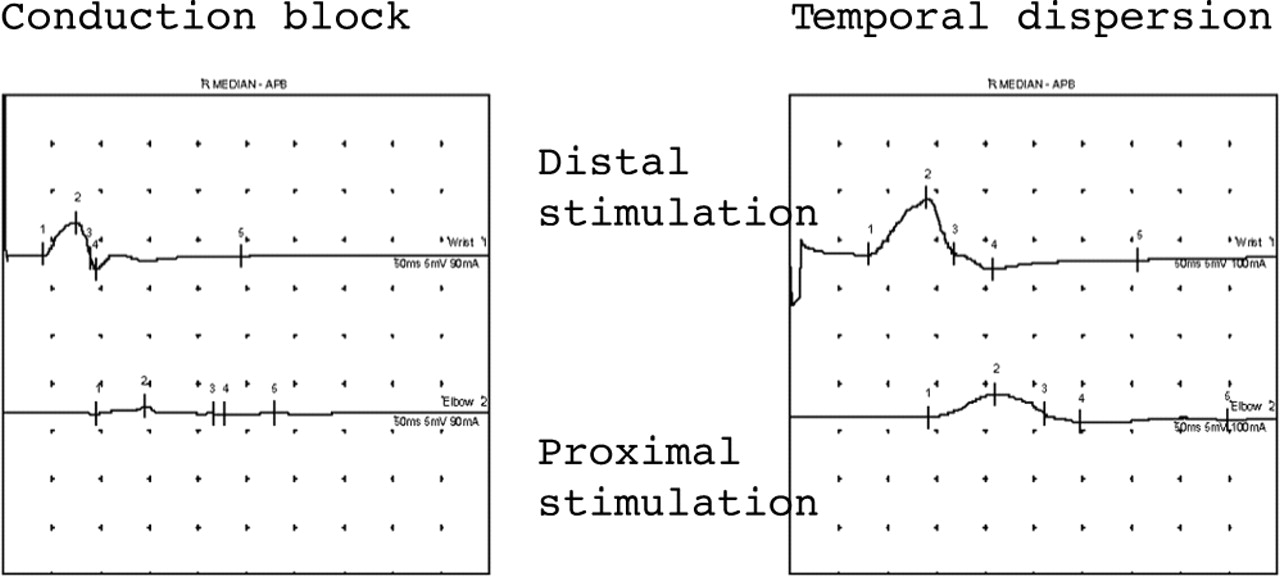
Both these traces show demyelination in median motor studies. The trace on the left shows almost complete conduction block with an absent response with proximal stimulation. The trace on the right shows temporal dispersion where the CMAP duration increases by almost 40% with proximal stimulation. In both situations the CMAP amplitude with proximal stimulation is smaller.
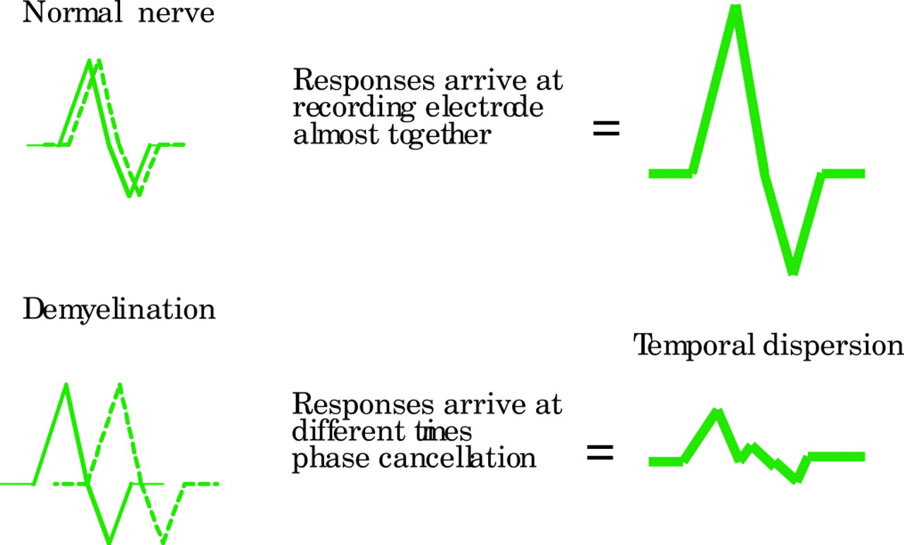
Schematic representation of phase cancellation and temporal dispersion in demyelination. In the normal nerve, the responses are synchronised in time and therefore summate (amplitude is higher that that of the individual components). Temporal dispersion results in an increased duration and reduced amplitude of CMAP.
Clinical correlates of motor conduction velocity slowing and conduction block
It is important to realise that slowing of conduction velocity alone without conduction block does not result in weakness as the impulses are still conducted from nerve to muscle. A good example of this is the presence of profound slowing of motor nerve conduction in totally asymptomatic primary relatives of patients with demyelinating hereditary motor and sensory neuropathy.
Sensory NCS
In generalised disorders
In both axonal and demyelinating pathologies the SNAP amplitude is reduced for different reasons. Sensory axonal loss will result in a smaller SNAP. Demyelination also produces small SNAPs but with prolonged durations. As they are of much shorter duration than CMAPs they are more susceptible to phase cancellation (fig 5).
The distribution of sensory NCS abnormalities may be helpful in determining aetiology. For example, the loss of SNAPS in the lower limbs is common in an axonal dying back neuropathy related to drugs like vincristine, whereas equal involvement of upper and lower limb SNAPS raise the possibility of a sensory ganglionopathy such as that related to thalidomide treatment.
Focal lesions
Multiple sensory NCS allow the investigator to locate sensory neuropathies that involve single or multiple digital nerves distally (for example, vasculitis or hand arm vibration syndrome) right up to the major trunks, cords, and divisions of the brachial plexus proximally.
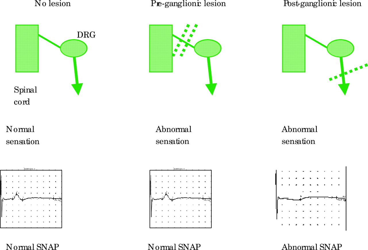
In proximal nerve trauma, maintenance of the sensory potential depends on the intact cell bodies in the dorsal root ganglia. Thus sensory NCS are extremely useful in localising a PNS lesion as either pre- and/or post-ganglionic. In a patient with a clinically suspected C8, T1 root lesion and with appropriate anaesthesia in that dermatome, the absence of the ulnar and medial antebrachial cutaneous sensory potential places the lesion distal to the dorsal root ganglion (DRG) in the lower trunk of the brachial plexus and not at root level (fig 6). Needle EMG can then be used to define this further.

Sensory responses are normal in pre-ganglionic lesions even though sensation may be abnormal clinically. Post-ganglionic lesions result in abnormal sensory responses. DRG, dorsal root ganglion.
F waves
Generalised disorders
F waves are sensitive to all forms of generalised peripheral neuropathy with their absence or a prolonged minimum latency occurring early. For example, in AIDP where demyelination may be segmental, proximal and patchy, F wave abnormalities may be the earliest and (in mild cases) the only electrophysiological abnormality seen.
In axonal pathology F wave latencies may also be mildly delayed in keeping with the motor conduction velocity slowing secondary to the loss of the fastest conducting motor axons.
In motor neuronopathies such as the motor neurone diseases, prolongation of any F wave latency is strong evidence either that this is the incorrect diagnosis (such as in multifocal motor neuropathy) or that a second pathological process is present.
Focal lesions
F waves may be absent in focal peripheral nerve or anterior spinal disorders. They were initially also thought to be very useful in identifying individual root distribution abnormalities. However, particularly in the upper limbs, the substantial overlap of segmental innervation in the distally available peripheral nerves makes this test on its own of low sensitivity and anatomical specificity. In addition, the effect of demyelination is diluted by the length of the path over which the F wave passes. In distinguishing the presence of a distal or proximal lesion, the use of the F wave ratio which compares the F wave latency in the upper and lower halves of the limb (conventionally using knee and elbow as the dividing line) may be useful.
REPETITIVE NERVE STIMULATION
Repetitive nerve stimulation (RNS) is used in the evaluation of patients with suspected neuromuscular transmission disorders (NMTD) such as myasthenia gravis (MG) or Lambert-Eaton myasthenic syndrome (LEMS). RNS is a modified motor NCS where instead of recording CMAPs with single supramaximal electrical stimuli, a train of 8–10 stimuli is applied and the sequential response amplitudes and/or areas measured. This may be carried out at low (3–4 Hz) or high frequency stimulation (20–50 Hz). In the latter case the train is prolonged to allow 2–10 seconds of continuous data to be measured. Both distal and proximal muscles/nerves should be studied in every patient suspected of an NMTD as the sensitivity of the test is greatly increased by this means.
With low frequency stimulation in normal subjects, the CMAP amplitude and/or area falls over the first 4–5 stimuli by a maximum of 10–12%. The maximum fall should be between potentials 1 and 2 (see RNS pitfalls). A number of department specific protocols have been published to study the RNS over time both before and after a period of maximum voluntary contraction of the muscle to pick up early or late NMT failure (fig 7).
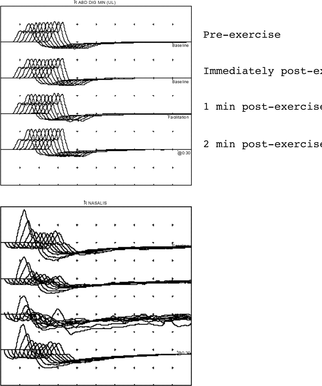
(A) Normal repetitive nerve stimulation from abductor digiti minimi muscle in the hand. The amplitude of the CMAPs within each train does not decrement nor is there any significant increment in CMAP amplitude after exercise. (B) Repetitive nerve stimulation in myasthenia gravis from the nasalis muscle. Four stimulus trains are given—all at baseline with no exercise. A reproducible decrement of 55% is seen (train 3 is technically unsatisfactory because of movement artefact).
High frequency stimulation may be used to discover evidence of a post-synaptic transmitter release disorder like LEMS. It is painful and requires considerable patient tolerance. There is evidence that recording low frequency RNS immediately before and after a 20–30 second period of maximum voluntary contraction by the patient is equally sensitive and is more humane (fig 8).

These traces show typical electrophysiological features of a pre-synaptic neuromuscular transmission disorder in a patient with LEMS. The traces on the left show a small amplitude ulnar CMAP that after exercise increases fourfold in amplitude. The traces on the right show repetitive nerve stimulation studies. The amplitude increases post-exercise. Trains 1, 3, and 4 show a decrement of 30% which repairs with exercise (train 2).
There are many pitfalls in the RNS test and artefact almost always gives rise to an abnormal test. Thus adherence to a strict protocol and heightened suspicion on the part of the CN to an abnormal result is essential as are repeated studies for reproducibility of abnormalities (see RNS pitfalls).
Physiological basis for the RNS
The neuromuscular junction consists of the motor axon terminal, the synaptic cleft, and the post-synaptic muscle membrane. As the motor axon potential depolarises the nerve terminal, voltage gated calcium channels open increasing the concentration of calcium in the pre-synaptic nerve terminal. This in turn facilitates the release of quanta of acetylcholine (ACh) from the nerve terminal into the synaptic cleft. ACh binds to receptors on the post-synaptic membrane causing depolarisation (end plate potential). The size of the end plate potential is dependent on the amount of ACh released and its binding to receptors. In the healthy state, the end plate potential reaches a threshold level and causes an action potential to be propagated along a muscle fibre resulting in muscle contraction. Normally there is a large safety factor for neuromuscular transmission with the amount of ACh released per impulse several times that required to generate a threshold level end plate potential.
In low frequency RNS, the rate of stimulation is such that the end plate physiology is stressed, but not to the level that produces the natural facilitation of NMT at greater stimulation frequencies. Thus an abnormal fall (decrement) in CMAP amplitude and/or area at low stimulation rates indicates a drop in the safety factor for transmission whether from a pre- or post-synaptic cause.
In high frequency stimulation natural facilitation is enhanced by pre-synaptic Ca++ influx and this may counteract a process such as LEMS where quantal release is depressed.
RNS in disease
NMT disorders may be congenital or acquired and in broad terms can be thought of as pre-synaptic or post-synaptic depending on where the defect lies.
Post-synaptic disorders of neuromuscular junction transmission
The archetypal post-synaptic disorder is myasthenia gravis (MG) where antibodies to acetylcholine receptors (AChR) cause degradation and increased turnover of receptor as well as macrophage initiated post-synaptic membrane simplification. In MG the safety factor is lost because as AChRs are depleted, less post-synaptic depolarisation occurs and some end plate potentials do not reach threshold for genesis of a propagated muscle membrane potential producing neuromuscular block. If this process affects a significant proportion of the tested muscle end plates, the RNS baseline train will show a significant decrement (greater than 10%). The decrement is usually measured by comparing the amplitude of the third or fourth CMAP in the train to the first (fig 7B). Very often some slight facilitation reduces the decrement over potentials 7–10 of the train.
An abnormal decrementing RNS test is non-specific and can be seen in a number of circumstances where muscle contraction processes may fail with repetitive stimulation (see RNS pitfalls).
Pre-synaptic disorders
In LEMS there are antibodies to voltage gated calcium channels (pre-synaptic disorder) causing impaired release of ACh quanta. Low frequency RNS stimulation may produce exactly the same decrement as seen in MG with additionally a small initial CMAP amplitude. Here calcium influx into the nerve terminal is reduced due to the action of voltage gated calcium channel antibodies and in turn ACh release into the synaptic cleft is reduced and some end plate potentials will be sub-threshold.
However, the diagnostic abnormality is of a significant > 100% increment in the CMAP amplitude after exercise or fast repetitive stimulation. Exercise increases calcium influx and the CMAP amplitude may increase by up to 10 times. In this case we are just comparing the amplitude of the first CMAP in the train before and after exercise (fig 8). Despite this increment, within each low frequency train a further decrement may occur due to ACh depletion.
PITFALLS
There are many pitfalls that can trap the unwary both in the performance and the interpretation of the NCS and RNS. For convenience these are separated in tables 2 and 3. The technical pitfalls more appropriately addressed to the reader who is an expert or training in CN are not included.
Performance of tests
Interpretation
CONCLUSIONS
Nerve conduction studies as part of the PNE are an extension of the clinical history and examination and are important in the management of cranial and peripheral neuromuscular disease as well as contributing to diagnosis of spinal cord lesions. NCS can be extremely useful both in localising lesions and determining the pathological processes responsible. We have listed many of the pitfalls both for the CN carrying out and interpreting the tests as well as for the referring doctor. For the former it is vital both to carry out tests accurately and reproducibly and to develop an investigation strategy based on the patient’s symptoms and signs rather than a fixed protocol. The investigator should then report the results clearly and then place them in the context of the clinical situation.
For the neurologist or other referring doctor, it is equally vital that the clinical questions asked are explicit and answerable for the most to be gained from what can be a considerable investment in time and skills for the investigator and tolerance of discomfort in the patient. For the best use of scarce resources therefore training and awareness of all the techniques detailed in this monograph are essential as part of general neurological training.