Article Text

Abstract
Background Delayed umbilical cord clamping (DCC) affects the cardiopulmonary transition and blood volume in neonates immediately after birth. However, little is known of blood flow in the umbilical vessels immediately after birth during DCC. The objective is to describe the duration and patterns of blood flow through the umbilical vessels during DCC.
Methods Arterial and venous umbilical blood flow was measured during DCC using Doppler ultrasound in uncomplicated term vaginal deliveries. Immediately after birth, the probe was placed in the middle of the umbilical cord, pattern and duration of flow in vein and arteries were evaluated until cord clamping.
Results Thirty infants were studied. Venous flow: In 10% no flow was present, in 57% flow stopped at 4:34 (3:03–7:31) (median (IQR) min:sec) after birth, before the cord was clamped. In 33%, flow continued until cord clamping at 5:13 (2:56–9:15) min:sec. Initially, venous flow was intermittent, increasing markedly during large breaths or stopping and reversing during crying, but then became continuous. Arterial flow: In 17% no flow was present, in 40% flow stopped at 4:22 (2:29–7:17) min:sec, while cord pulsations were still palpable. In 43% flow continued until the cord was clamped at 5:16 (3:32–10:10) min:sec. Arterial flow was pulsatile, unidirectional towards placenta or bidirectional to/from placenta. In 40% flow became continuous towards placenta later on.
Conclusions During delayed umbilical cord clamping, venous and arterial umbilical flow occurs for longer than previously described. Net placental transfusion is probably the result of several factors of which breathing could play a major role. Umbilical flow is unrelated to cessation of pulsations.
- birth
- cord clamping
- umbilical flow
Statistics from Altmetric.com
What is already known on this topic
-
Delayed umbilical cord clamping is beneficial for cardiopulmonary transition and blood volume in infants at birth.
-
Delayed cord clamping is now recommended, but there is a variation in the advised time point and in some guidelines no maximum time is recommended.
-
Understanding the physiology of umbilical flow after birth can be helpful to understand the benefits/consequences of delayed cord clamping.
What this study adds
-
During delayed cord clamping venous and arterial umbilical flow occurs for longer than previously described.
-
Placental transfusion is complex and several factors, including breathing, and whether venous and/or arterial flow is still present, has to be taken into account.
-
Umbilical flow is unrelated to cessation of pulsations, and using this as a time point for cord clamping should be reconsidered.
Background
Until 1960, allowing time for placental transfusion by delaying cord clamping (DCC) was common and advocated by famous caregivers over the centuries.1 ,2 With improved medical care and more women giving birth in hospitals, early/immediate cord clamping was introduced as an intervention to reduce the risk of postpartum haemorrhage with no clear reason.2 The practice of cord clamping has changed over recent decades, but remains a controversial subject with numerous publications and meta-analysis discussing the advantages and disadvantages of early or delayed cord clamping.3 ,4 Several guidelines and experts now recommend delayed cord clamping, but there is a variation in the advised time after birth and in some guidelines no maximum time is recommended.4
Despite the debate on DCC, little is known about the factors regulating blood flow in the umbilical vessels immediately after birth. The concept of net placental-to-infant-transfusion during DCC has been studied indirectly by measuring circulating blood volume,5 the residual placental blood volume6 and more recently by the change in infant weight while the cord is intact.7 The contribution of the umbilical arteries was considered to be minimal as it was thought that arterial flow stopped within 25–45 s.8 However, no explanation for this cessation of flow was provided, despite the fact that umbilical arterial flow can continue for hours in exteriorised fetuses, as long as the fetal state is maintained.9 In addition, recent animal data indicate that for a stable haemodynamic transition any delay should not be time-based, but should depend on the infant's physiology, particularly whether the infant is breathing or not.10 In addition, onset of breathing and effort would also influence placental transfusion.11–13
To understand the physiological benefits/consequences of DCC, more knowledge about the physiology of umbilical flow after birth is important. Non-invasive techniques such as ultrasound can be easily used to monitor umbilical flow after birth and are well tolerated by the infant and mother. We therefore investigated the duration and pattern of umbilical venous and arterial flow in the umbilical cord of healthy term infants directly after birth using ultrasound.
Methods
We conducted a prospective, observational, pilot study using ultrasound to assess the flow patterns in the umbilical cord vessels directly after vaginal birth at term. The study was performed in the Leiden University Medical Center by a researcher who was trained and supervised by a neonatologist and a cardiologist. During 3 months we approached women with singleton pregnancies, >37 weeks gestation for participation in this study. Exclusion criteria were pre-eclampsia, antihypertensive or anticoagulant drugs or detected congenital disorders or intrauterine growth retardation (fetal biometry with estimated fetal weight and abdominal circumference below the 2.3th centile). No infants received respiratory support immediately after birth and infants from multigravida and primigravida pregnancies were eligible.
The study was approved by the institutional review boards of the Leiden University Medical Center (Commissie Medische Ethiek, Leids Universitair Medisch Centrum) and informed consent was obtained on the day of delivery, or earlier, but before the women were in active labour.
Direct skin-to-skin contact is standard of care and infants were placed on the mother's chest directly after birth. Ultrasound of the umbilical cord did not interfere with the care of either the mother or baby. The management of third stage of labour, including cord clamping, was carried out at the midwives’ discretion. Although not governed by local guidelines, DCC is common in our hospital, where the cord is clamped at least 1 min after delivery of the baby or when pulsations have ceased.14
As soon as the infant was placed on the mother’s chest an ultrasound probe was gently placed on a straight section of the umbilical cord to visualise all vessels. The midwife ensured the umbilical cord was positioned in an easily accessible spot for the researcher. Measurements continued until the cord was clamped, the timing of which was determined by the midwife. The researcher examined the umbilical cord to detect for pulsation by palpation immediately before cord clamping, to relate the presence or absence of pulsations to the measured umbilical blood flow.
Recordings were performed using a Vivid-I (GE Healthcare) ultrasound machine and a neonatal/paediatric 10 S (4–10 MHz) probe. The probe was placed gently on the umbilical cord and aligned with the direction of blood flow at an angle of <20°. The umbilical arteries and vein were detected, then using colour Doppler the flow direction was determined and, using M-mode the pulsatility was assessed. The velocity scale was set as low as possible (0.3–0.4 m/s). No flow was defined as no visual colour on colour Doppler, meaning little to no flow in the umbilical artery and vein. During the recordings the probe was held steady. Recordings were saved for off-line analysis. In most instances, it was only possible to determine the direction and pattern of blood flow in one vessel at a time. As we could not guarantee a consistent angle of the probe and location during the period of measurements, we were not able to measure blood volume. The duration of flow phenomena were measured from start of colour flow until the start of the next colour flow and frequencies were calculated from this. Therefore, we recorded blood flow (colour Doppler+M-mode) for 15 s alternately in the vein and in one of the arteries. During off-line analysis (EchoPac GE), the presence and direction of flow and pulsatility of flow were related to the time after birth and the time of cord clamping.
The following basic parameters were also recorded: gestational age, parity, birth weight, sex, time from birth (when the first shoulder was out) to cord clamping, time of the first breath, whether the infant was crying at birth, time the placenta was delivered, and when the midwife injected uterotonics, Apgar score, vaginal blood loss and weight of the placenta. To accurately record all time points, a multitrack stopwatch was used.
Outcome variables
The flow patterns in the arteries and veins were described; whether they were pulsatile, continuous, unidirectional or bidirectional and when this occurred. The effect of breathing and crying on the flow and patterns were observed and described. The time after birth at which the arterial and venous blood flow stopped was noted. It was noted whether cord pulsations were present after the flow stopped or right before cord clamping when flow was still present.
Statistical analysis
Data is presented as median (IQR), mean±SD or number (percentage) where appropriate. Time points are expressed in min:sec after birth. For database and statistical analysis SPSS software V.20.0 (SPSS, Chicago, Illinois, USA; 2012) was used.
Results
Consent to perform the measurements was given by 43 women. Thirteen cases were excluded: in eight infants the researcher arrived too late, one needed resuscitation and four were delivered by a secondary caesarean section. Thus measurements were made in 30 infants (table 1). Oxytocin (5 IU) was given immediately after birth to 28 mothers. Most infants took their first breath directly after birth (table 1). In all infants the first measurements were made after the first breath and after administration of oxytocin.
Infant characteristics and time points
Venous flow
Timing
In 3/30 (10%) infants absence of venous flow was observed at the initial examination. In 17/30 (57%) venous flow stopped at 04:36 (03:03–08:22) min:sec after birth and the cord was clamped 06:02 (04:47–09:35) min:sec after birth. In 10/30 (33%) venous flow was still present when the cord was clamped at 05:13 (02:56–09:15) min:sec after birth.
Flow pattern
In the 27 (90%) infants, where venous flow was observed, it was initially intermittent during the first breaths. During large breaths the venous flow increased (figure 1) during inspiration, but the flow completely stopped when the infant cried during expiration (figure 2) or flow even reversed when crying hard (observed in 7/27 (26%); figure 2). The frequency of this phenomenon ranged from 20/min to 50/min, which most likely reflected the respiratory rate of the infants. With time, when breathing was more regular and less vigorous, the venous flow became more continuous and decreased until it stopped.
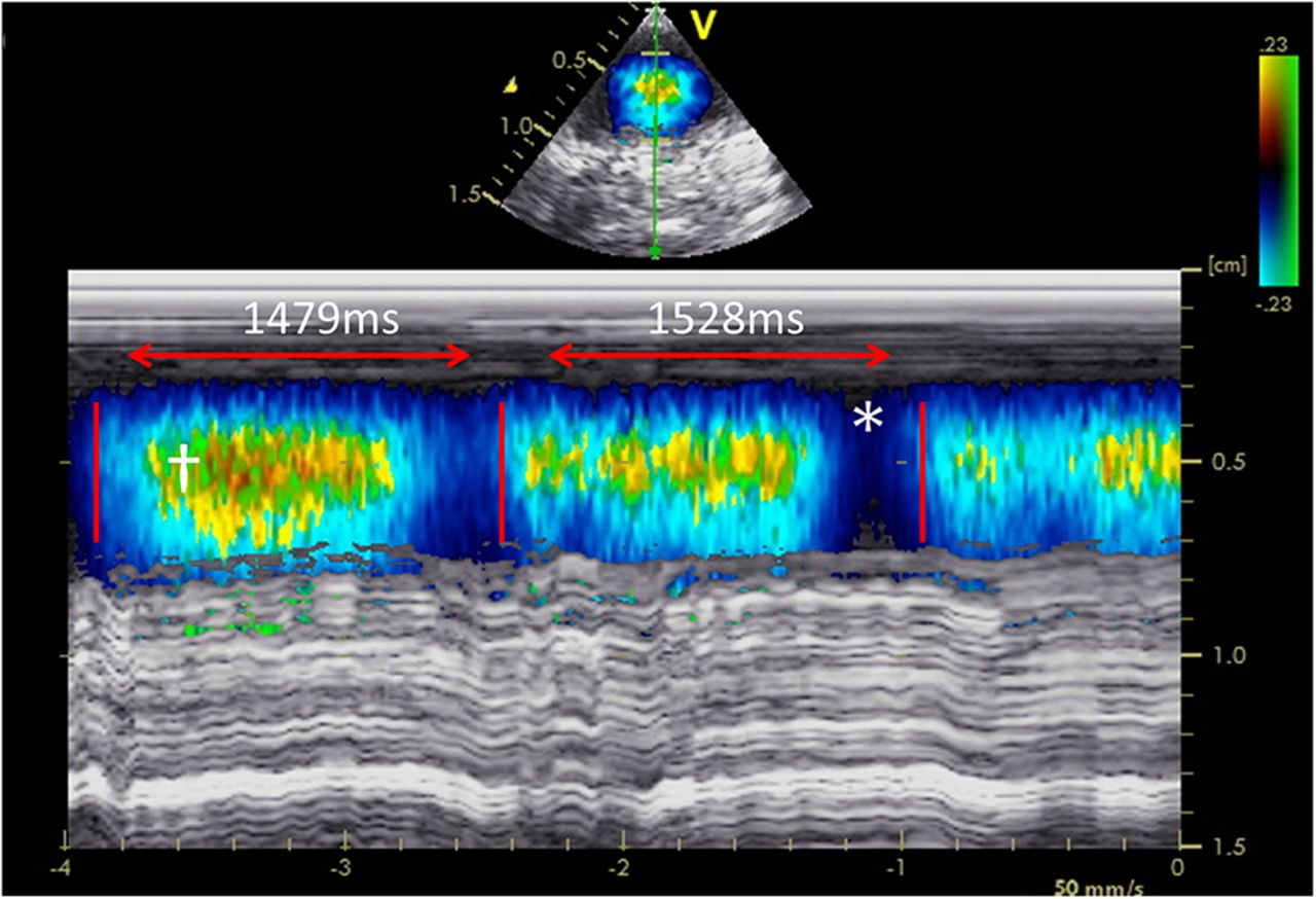
Example of a flow pattern in an umbilical vein during breathing. There is flow (blue, direction towards infant) with an increasing velocity creating aliasing of the Doppler flow signal (†) after which it decreases. In the middle, flow stops for a brief moment (*). Time from start of flow until start of next flow pattern is ∼1500 ms, which may reflect a breathing rate of ∼40/min.

Example of a flow pattern in an umbilical vein during crying. There is short period of flow (blue, direction towards infant) with an increasing and decreasing velocity (during the short large inspiration from the cry17) after which flow stopped during the expiration of the cry17 with a short period of reversed flow (red, direction to placenta) in the middle. Time from start of flow towards the infant until the start of the next flow pattern was ∼2103 ms, which may reflect a breathing rate of ∼28/min.
Arterial flow
Timing
In 5/30 (17%) infants absence of arterial flow was observed from the start of initial measurement until cord clamping at 04:45 (02:45–07:45) min:sec after birth. In 12/30 (40%) infants arterial flow was observed but stopped 04:22 (02:29–07:17) min:sec after birth before the cord was clamped at 06:15 (05:02–09:30) min:sec. In 11 of these 12 infants pulsations were still palpable after the flow detected by ultrasound had stopped (including venous flow). In 13/30 (43%) arterial flow was still detectable when the midwife decided to clamp the cord at 05:16 (03:32–10:10) min:sec after birth.
Flow pattern
In the 25/30 (83%) infants where arterial flow was observed, flow was pulsatile, occurring at a pulse rate similar to the infant's heart rate and flowed from the infant to the placenta. The flow was mainly unidirectional (flow towards the placenta during systole, with no flow during diastole). However, in 18/25 (72%) an intermittent bidirectional flow (towards the placenta during systole, and away from the placenta during diastole) was observed (figure 3). The frequency of this phenomenon ranged from 120/min to 180/min, which most likely reflects the heart rate of the infants. This bidirectional flow appeared 00:45 (00:25–02:36) min:sec after birth and the median duration was 02:24 (01:37–03:52) min:sec. In 10/25 (40%) infants, the pulsatile arterial flow eventually ceased and became continuous flow from the infant to the placenta.

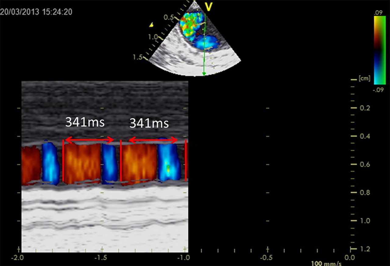
Example of a pulsatile, bidirectional flow pattern in an umbilical artery. Red is towards the placenta and blue towards the infant. The duration of one pattern is ∼341 ms, which may reflect the infant's heart rate of 175/min.
Time difference venous-arterial
In 15 infants venous and arterial flow stopped at the same time, either before cord clamping or at the time of cord clamping. In seven infants arterial flow stopped before venous flow with a time difference of 01:08 (00:51–03:03) min:sec. In eight infants, venous flow stopped before arterial flow with a time difference of −01:43 (−00:51–02:45) min:sec.
Discussion
This is the first study to directly investigate umbilical cord flow immediately after birth during delayed cord clamping. We observed that umbilical venous and arterial blood flows continue longer than previously described.6 ,8 The duration of flow is however, highly variable between subjects. The observation that arterial flow can continue after venous flow has stopped (a third of the infants) has not been described before and might influence net placenta-to-infant transfusion. Loss of umbilical cord pulsations was not correlated to the actual arterial flow. The large influence of the infants’ first breaths, including crying, on the venous flow patterns suggests that breathing could play an important role in the net placental transfusion. Although it was not possible to quantify the arterial and venous blood volume changes, the findings in this study imply that postnatal placental transfusion is more complicated than currently assumed.
Yao et al,5 indirectly measured the net placental transfusion by measuring placental residual blood volume and neonatal blood volume using the 125I-labelled albumin dilution technique, which relies on calculations extrapolated from adult studies. They drained the placenta for 20 min after cord clamping and observed very little residual volume when cord was clamped at 3 min,5 which is now the current prevailing view in the literature.15 We observed that arterial and venous flow can occur for a much longer duration than previously envisaged, but it is debateable as to how much placenta-to-infant transfusion occurs if cord clamping is delayed for these periods. It is possible that net transfusion is rapid, depending upon the compliance and pressure gradients between the two circulations, achieving a balance whereby no further net transfusion occurs. However, in a large proportion of the infants venous and arterial flow stopped at different time points, which likely resulted in a net gain or loss in transfusion volume. Farrar et al7 weighed the infants while cords were intact and observed that in some infants weight continued to increase after 3 min, which is consistent with our observations.
The observation that breathing influenced the flow pattern in the umbilical vein is consistent with previous studies reporting that the onset of breathing and the degree of effort influences placental transfusion.11–13 During inspiration umbilical venous flow towards the infant increased, which was reduced or ceased during expiration. It is likely that the large subatmospheric intrathoracic pressures generated by the first inspiratory efforts increase the placental-atrial pressure differences favouring flow into the infant.16 Crying is essentially a forced expiration against a partially closed glottis, leading to supra-atmospheric pressure in the lung17 and probably caused umbilical venous flow to cease or reverse towards the placenta. It was recently demonstrated in preterm lambs that ventilation before cord clamping stabilised the haemodynamic transition at birth by increasing pulmonary blood flow before umbilical venous return was lost.10 However, in that experiment, positive pressure ventilation was used which would markedly alter the placental-atrial pressure gradient compared with spontaneous breathing, possibly indicating that benefits of DCC may be enhanced in spontaneously breathing infants.
In our study arterial flow continued much longer than described in a study where residual placental blood volumes were compared when arteries and veins were clamped separately at various times.8 The differences in methodology probably explain the contradictory findings. The left to right ductal shunting caused by a decrease in pulmonary vascular resistance and the increase in placental vascular resistance by uterine contractions could explain the absent diastolic flow or bidirectional flow we observed. The more continuous and much less pulsatile flow later on might reflect an increase in placental vascular compliance when detachment occurred. We did not record ductal blood flow or contractions of the uterus and so further observations are needed to confirm or refute this.
Although no data are available, and the rationale is uncertain, the cessation of umbilical cord pulsations has been used as time point for cord clamping, probably based on the belief that flow stops when pulsations stop.4 In our study the presence or absence of pulsations appeared unrelated with flow in the venous and arterial umbilical vessels, indicating that cardiac generated pressure waves continue to reach the umbilical cord irrespective of whether arterial flow has ceased. This implies that using cessation of pulsation as a time point for cord clamping should be reconsidered.
Several factors (timing, delivery mode, presence of intrapartum asphyxia, breathing, gravity and uterine contractions) could influence the net placenta-to-infant transfusion immediately after birth.5 ,11–13 ,18–22 We only report infants born vaginally, who breathed immediately and were placed above the placenta, and all mothers received oxytocin immediately after delivery. It is possible that change in any of these factors would lead to different observations.20 ,21 However, a recent trial has demonstrated that gravity had very little influence on the placental transfusion.23 It is of interest that Yao et al21 observed that placental transfusion was complete within a minute when ergometrine was given immediately after birth. Ergometrine is a more powerful uterotonic than oxytocin, which might explain that in our study arterial and venous flow was still present minutes after birth in most infants.
Although the sample size was small, we have shown that it is feasible to make ultrasound recordings of umbilical flow during DCC, from which we have made unique observations. We were not able to reliably quantify the flow as we could not guarantee a consistent angle and location of the probe on the umbilical cord. Currently other ways to measure reliable flow are being investigated. Another limitation was that it was not possible to observe flow in the vein and arteries simultaneously, forcing us to alternate between the vein and artery every 15 s. As such, our observations on the timing of the cessation of flow could be in error by a maximum of 15 s in any individual baby. It is possible that flow was influenced by manipulating or compression of the cord by the ultrasound probe. However, we minimised this by placing the probe very gently on the cord.
In summary, this study provided unique observations of umbilical cord blood flow directly after birth, before cord clamping. The flow patterns observed were in contrast to current concepts and in many babies flow does not stop within 3 min. The mechanism of placental to infant blood transfusion is complex and several factors, including breathing, particularly crying, and whether venous and/or arterial flow are still present, have to be taken into account for the net effect of DCC. For a recommendation concerning the timing of cord clamping, and also if there is a minimum and maximum duration, further studies are needed to understand the physiology of DCC.
References
Footnotes
-
Contributors IB performed the data collection, carried out the analyses, drafted the initial manuscript and approved the final manuscript as submitted. AAWR contributed to the design of the study, supervised the data collection and carried out the analyses, reviewed and revised the manuscript, and approved the final manuscript as submitted. EW, ADJtH and MCH reviewed and revised the manuscript, and approved the final manuscript as submitted. CJM and SBH conceptualised the study, reviewed and revised the manuscript, and approved the final manuscript as submitted. ABtP conceptualised and designed the study, supervised the data collection and analysis, reviewed and revised the manuscript, and approved the final manuscript as submitted.
-
Funding ABtP is recipient of a Veni-grant, The Netherlands Organisation for Health Research and Development (ZonMw), part of the Innovational Research Incentives Scheme Veni-Vidi-Vici, project number 91612027.
-
Competing interests None.
-
Patient consent Obtained.
-
Ethics approval The Institutional Review Boards of the Leiden University Medical Center (Commissie Medische Ethiek, Leids Universitair Medisch Centrum).
-
Provenance and peer review Not commissioned; externally peer reviewed.
Linked Articles
- Fantoms