Article Text

Abstract
Introduction Cognitive impairment and reduced well-being are common manifestations of Graves’ disease (GD). These symptoms are not only prevalent during the active phase of the disease but also often prevail for a long time after hyperthyroidism is considered cured. The pathogenic mechanisms involved in these brain-derived symptoms are currently unknown. The overall aim of the CogThy study is to identify the mechanism behind cognitive impairment to be able to recognise GD patients at risk.
Methods and analysis The study is a longitudinal, single-centre, case-controlled study conducted in Göteborg, Sweden on premenopausal women with newly diagnosed GD. The subjects are examined: at referral, at inclusion and then every 3.25 months until 15 months. Examinations include: laboratory measurements; eye evaluation; neuropsychiatric and neuropsychological testing; structural MRI of the whole brain, orbits and medial temporal lobe structures; functional near-infrared spectroscopy of the cerebral prefrontal cortex and self-assessed quality of life questionnaires. The primary outcome measure is the change in medial temporal lobe structure volume. Secondary outcome measures include neuropsychological, neuropsychiatric, hormonal and autoantibody variables. The study opened for inclusion in September 2012 and close for inclusion in October 2019. It will provide novel information on the effect of GD on medial temporal lobe structures and cerebral cortex functionality as well as whether these changes are associated with cognitive and affective impairment, hormonal levels and/or autoantibody levels. It should lead to a broader understanding of the underlying pathogenesis and future treatment perspectives.
Ethics and dissemination The study has been reviewed and approved by the Regional Ethical Review Board in Göteborg, Sweden. The results will be actively disseminated through peer-reviewed journals, national and international conference presentations and among patient organisations after an appropriate embargo time.
Trial registration number 44321 at the public project database for research and development in Västra Götaland County, Sweden (https://www.researchweb.org/is/vgr/project/44321).
- Graves' disease
- hippocampus
- cognitive impairment
- magnetic resonance volumetry
- functional near-infrared spectroscopy
- quality of life
This is an open access article distributed in accordance with the Creative Commons Attribution Non Commercial (CC BY-NC 4.0) license, which permits others to distribute, remix, adapt, build upon this work non-commercially, and license their derivative works on different terms, provided the original work is properly cited, appropriate credit is given, any changes made indicated, and the use is non-commercial. See: http://creativecommons.org/licenses/by-nc/4.0/.
Statistics from Altmetric.com
- Graves' disease
- hippocampus
- cognitive impairment
- magnetic resonance volumetry
- functional near-infrared spectroscopy
- quality of life
Strengths and limitations of this study
Prospective case-controlled study.
Extensive neuropsychological and neuropsychiatric assessment.
State-of-the-art manual and automatic analysis of structural magnetic resonance images of medial temporal lobe structures.
Use of a novel functional technique, functional near-infrared spectroscopy, to determine correlates of mental fatigue.
A limitation is that only women are studied.
Introduction
Cognitive dysfunction is a common late complication of Graves’ disease (GD), but the pathogenic mechanisms involved are currently unknown. Advanced imaging methods that provide new perspectives into the central nervous system are being increasingly used for functional and structural studies of neurological and psychiatric disorders. The CogThy project is investigating neural correlates of GD in a longitudinal study to expand understanding of the underlying pathogenic mechanisms of brain involvement in the disease.
GD: a disease with immune system dysregulation
GD is the most common form of hyperthyroidism in Sweden with an incidence of 21 per 100 000 inhabitants.1 The lifetime risk is 3% for women and 0.5% for men.2 In GD, stimulating autoantibodies directed against the thyroid-stimulating hormone (TSH) receptor result in elevated levels of circulating thyroid hormones (thyroxine (T4) and tri-iodothyronine (T3)) and reduced levels of circulating TSH. The high thyroid hormone levels affect most organ systems through genomic pathways, although there are also non-genomic effects of T3 on membrane receptors3 (eg, on mitochondria and on the receptors of sympathetic nerves.4
GD may occur at any age, but the peak incidence is between 30 and 50 years of age.2 5 Hence, it often affects people of working age and leads, in many cases, to months of lost productivity.6 Moreover, GD may be complicated by extrathyroidal autoimmune manifestations, such as thyroid-associated ophthalmopathy (TAO) with an incidence of 4.9%,7 thyroid dermopathy occurring in 1%–4% of patients and acropachy (swelling of the hands and clubbing of the fingers).2
Thyroid hormones affect brain function
Thyroid hormones are essential for normal brain development and adult brain function. Substantial iodine deficiency during critical periods of brain development results in hypothyroidism, leading to severe and irreversible cognitive, neurological and IQ impairments.8 Acquired adult overt hypothyroidism also entails mental signs and symptoms with reduced information processing speed, reduced efficiency of executive functions and learning difficulties. Furthermore, hypothyroidism is associated with an increased risk of depression and lower health-related quality of life (QoL). These symptoms generally improve with levothyroxine replacement treatment.9
Conversely, in hyperthyroidism, the psychiatric symptoms are often striking with unrest, stress intolerance, fatigue, memory impairment and compromised well-being.10–12 Patients may also experience anxiety and depression,13 and cognitive impairment.14
Psychiatric signs and symptoms in GD
Many studies indicate that hyperthyroid patients suffer from affective symptoms10–13 15–21 and some provide evidence for hyperthyroidism being associated with cognitive dysfunction.10 15 19 22 23 The main cognitive dysfunction seen in patients with GD may be described as an astheno-emotional disorder,24 25 which is characterised by difficulty in maintaining attention during cognitively demanding tasks, with a consequent tendency to develop fatigue, memory difficulties, irritability and emotional lability. Episodes of secondary depression are often seen late in the course. Astheno-emotional disorder, often called mental fatigue,26 is present in many pathological conditions including traumatic brain injury, brain tumours, stroke27 and endocrine diseases such as Cushing's disease and GD. Possible alternative labels to astheno-emotional disorder/mental fatigue are mild cognitive disorder28 and mild neurocognitive disorder.29 However, descriptions of the latter two conditions do not emphasise the characteristic mental fatigability in astheno-emotional disorder/mental fatigue and, importantly, do not include emotional signs and symptoms.
Although euthyroidism is usually achieved rapidly under treatment, recovery of well-being is delayed for months in many patients, and some never regain full pre-morbid health.10 12 13 30 Approximately 20% of patients experience reduced QoL for as long as 1 year after treatment11 and many are not fully recovered after 3 years.16 A randomised, prospective study31 found patients who received treatment for GD with antithyroid drugs (ATDs), radioactive iodine or surgery still had decreased vital and mental QoL 17–21 years later compared with the general population. Several prospective studies10 11 13 32 in patients with GD indicate a short-term negative impact on QoL as measured by a 36-item short-form health survey and, more recently, a thyroid-specific questionnaire (ThyPRO).
Sick leave is more prevalent in GD patients 1 year after diagnosis than in the general population.16 33 Patients with hyperthyroidism have a doubled risk for being absent from work during the first year after start of treatment and fewer return to work even during subsequent years.6 These observations are in agreement with a previous report34 where more than 30% of patients reported complete or partial inability to work 6 years after GD diagnosis. It is unknown whether the dominant reason for the prolonged inability to return to full-time work is impaired mental health or somatic symptoms.
Thyroid hormones, thyroid antibodies and autoimmunity
Thyroid hormones, especially T4, are actively transported into the brain.35 Most of the active thyroid hormone, T3, is converted locally in the brain from T4 by deiodinase type 2 (D2),36 which regulates tissue-specific intracellular T3 production and thereby protects tissues from hypothyroidism and hyperthyroidism. D2 activity is lower in hyperthyroidism.37 T3 interacts with T3 receptors (TRs) distributed throughout most brain regions, with the highest levels found in the olfactory bulbs, the hippocampus and the cerebellar cortex.38–40 Two distinct genes, TRα and TRβ, encode several receptor isoforms with specific functions that define the tissue-specific action of thyroid hormone in the brain.41–43
Besides TR activation and non-genomic pathways of thyroid hormones, TSH receptors may be involved in the effects on the brain present in GD, as TSH receptors are stimulated by TSH-receptor autoantibody (TRAb). TSH receptors are seen in the hippocampi and in the neocortex,44 and their action is different from that of TRs.45 Moreover, the production of autoantibodies in GD is not limited to those directed against the TSH receptor. Autoantibodies against thyroglobulin, thyroperoxidase and insulin-like growth factor 1 receptor,46 angiotensin II receptor type 147 and β1-adrenergic and M2 muscarinic receptors48 have all been described as elevated in GD, with proposed cardiovascular pathogenic effects.47 48 Hence, brain pathology related to GD may be caused by intracerebral hyperthyroidism, thyroid-related antibodies and/or, more directly, the autoimmunity per se.49 Supporting the latter two alternatives is the fact that depression and anxiety appear more prevalent in GD than with toxic nodular goitre.13
Knowledge gaps and background to the CogThy study
Reduced grey matter volume of the hippocampus has been described in adult hypothyroidism using MRI.50 The mechanism behind this was thought to be related to a reduction in hippocampal neurogenesis, as evidenced by animal studies.51
Another study in adult hyperthyroidism has provided evidence that excess thyroid hormones influence brain function and lead to reduced hippocampal volume.52 In contrast, animal studies of hyperthyroidism found no effect on hippocampal progenitor cell proliferation, survival or differentiation.51 53 In elderly humans, higher T4, but within the normal range, is correlated with hippocampal and amygdalar atrophy,54 but no association has been identified between medial temporal brain volumes and polymorphisms of D2 that converts T4 to T3.55
Claims that medial temporal lobe (MTL) structures are implicated in cognitive impairment in GD are supported by observations in TRα1 knockout mice.56 These findings indicate that TRα1 is involved in the regulation of hippocampal structure and function, as the mice present considerable behavioural changes.56 Improved hippocampus-dependent learning and memory have been reported after T4 substitution in thyrectomised rats.57
Human studies on structural brain changes in hyperthyroidism have been cross-sectional. However, it is unknown whether structural and/or functional changes in the brain accompany the achieved euthyroidism and whether the brain's morphological changes are correlated with changes in cognitive functioning. Furthermore, it is largely unclear whether patients’ psychiatric diagnoses influence outcome and whether biological markers can be used to predict recovery from cognitive impairment. In addition, whether MTL structures, other than the hippocampus, are affected remains to be investigated. Finally, only a few GD studies17 22 58 have addressed the functioning of the frontal lobes, which are generally regarded as being crucial for cognition. The CogThy project aims to fill these gaps in knowledge.
Hypotheses
It was hypothesised that patients with GD exhibit reversible neuroplastic changes in the MTL and/or the prefrontal regions of the brain but the changes are persistent in those with sustained cognitive impairment.
Objectives
The primary objectives are to study structural and functional brain changes in patients with GD. Secondary objectives are to investigate neural correlates of neuropsychological, neuropsychiatric, hormonal and autoantibody findings, with the aim of being able to identify GD patients at risk of developing cognitive impairment and, subsequently, to adapt their treatment schedule accordingly.
Methods and analysis
Design of the CogThy study
The CogThy study is a prospective, longitudinal, case-controlled, observational study with an open treatment period for hyperthyroidism of ≥15 months in 65 female patients with GD and newly diagnosed hyperthyroidism. The reason for only including women is, in part, to minimise variables that might affect MTL volume, as there may be sex differences in these brain areas.59 In addition, there is also a more practical reason: both GD and affective disorders such as depression and anxiety are more common in women than in men. The patients undergo assessment at referral when ATDs are instituted and when hyperthyroidism develops. After inclusion, they are followed every 3.25 months and, finally, at 15 months, allowing for a period of euthyroidism ≥6 months (figure 1). Euthyroidism is defined as a serum TSH concentration between 0.4 and 2.0 mIU/L combined with normal free T4 (fT4) (12–22 pmol/L) and T3 (1.3–3.1 nmol/L) levels. Treatment of hyperthyroidism is in accordance with the local treatment scheme for non-elderly, female, adult patients with GD where all patients receive ATDs or surgery, in addition to previous ATD treatment, avoiding radioactive iodine treatment if possible. Hypothyroidism is prevented by starting T4 treatment early.

Study design. description of the time frame and assessments included at each visit for 65 patients and 65 controls included in the CogThy study.
The study visits at inclusion and at 15 months include somatic and eye evaluations, laboratory hormonal and antibody measurements, neuropsychological and neuropsychiatric evaluation, self-administered QoL questionnaires, magnetic resonance (MR) scanning of the brain (full coverage, and focal scanning of the MTL and orbits) and functional near-infrared spectroscopy (fNIRS). A 7.5-month evaluation includes only somatic and eye evaluations, laboratory measurements, full coverage MRI of the brain and focal MRI of the orbits. Evaluations at 3.25 and 10.75 months include only laboratory measurements (figure 1).
Patient and public involvement
For clinicians, mental fatigue is a well-known symptom among patients. Equally, patients are eager to understand the underlying pathogenic mechanism in brain symptoms. Patients were not involved in the design of the study but are involved by being recruited into the study. Participating patients will be informed about results by mail. Patient organisations will also be involved in the study results in due time.
Setting and participants
The study includes 65 consecutive, premenopausal, adult women with newly diagnosed GD presenting with fT4 ≥50 pmol/L (reference range: 12–22 pmol/L) and/or T3 ≥6.0 nmol/L (reference range: 1.3–3.1 nmol/L) at the Thyroid Unit of Sahlgrenska University Hospital, Göteborg or Kungälv Hospital, Kungälv, Sweden. The GD diagnosis is verified by positive TRAb and/or a homogeneous uptake on technetium scintigraphy. Inclusion and exclusion criteria are presented in box 1.
Inclusion and exclusion criteria
Inclusion criteria
Premenopausal women>18 years of age.
Newly diagnosed* GD.
fT4 ≥50 pmol/L (reference range: 12–22 pmol/L) and/or T3 ≥6 nmol/L (reference range: 1.3–3.1 nmol/L).
Positive TSH receptor antibodies and/or homogeneous uptake on technetium scintigraphy.
Exclusion criteria
Other concomitant illnesses affecting QoL such as other endocrine diagnoses, heart failure, respiratory insufficiency, active malignancy and current or recent psychosis diagnosis.
Patient who may not attend to the protocol according to the investigator’s opinion, for example, claustrophobia, planned emigration.
TAO with concomitant steroid medication or TAO where the use of steroids will be very likely within the next 15 months.
Concomitant or recent illness with steroid treatment.
Contraindications for MRI.
Amiodarone-induced GD.
fT4, free thyroxine; GD, Graves’ disease; QoL, quality of life; TAO, thyroid-associated ophthalmopathy; TSH, thyroid-stimulating hormone; T3, tri-iodothyronine.
*Newly diagnosed means that the general practitioner has just identified elevated fT4 and low TSH in serum tests and made a hospital referral. Blood tests are rechecked before the first visit and these lay the grounds for inclusion.
Control subjects with matching age and sex are selected from a random sample from the general population in Göteborg provided by the Swedish Tax Registry, who were contacted by regular mail. When a control subject has accepted participation, she is also matched regarding smoking status and educational level. In addition to the exclusion criteria stated for patients, controls are excluded if they have thyroid hormone levels outside the normal reference range, previous or ongoing thyroid disease, or TAO.
Inclusion
After referral to the hospital, eligible patients are approached either at or before the first clinical visit (which is when ATD treatment is started) to schedule an inclusion visit within 2 weeks after the first visit. In Sweden, ATD treatment is always prescribed by hospital endocrinologists. Earlier, the patient has often seen a general practitioner, who has prescribed beta-blocking agents and referred the patient. The time from referral to the first clinical visit is usually 1–2 weeks.
Dropouts
The sample size estimate allows for a substantial number of dropouts. The patients included are young or middle-aged women, some of whom will move away from the region or will not prioritise follow-up visits, even though the person recruiting the patient at inclusion carefully stresses the importance of attendance. Reasons for leaving the study are recorded, if known, and a sensitivity analysis will be performed to compare baseline data between dropouts and those included in the study.
Measures
The outcome measures are specified as follows. The primary outcome is the change in MTL brain volume. Secondary outcomes are regional cerebral blood flow, QoL, mental fatigue and other cognitive variables, psychiatric diagnoses, thyroid hormones, autoantibodies and D2 polymorphisms.
Assessment of background variables
Information is collected about ethnicity, socioeconomic status, use of alcohol and tobacco products, stress, pregnancy, family history of disease, previous low-dose irradiation to the head and neck region, head trauma, other previous and current diseases and ongoing medications.
Endocrine assessment
Blood specimens are analysed at the Department of Clinical Chemistry, Sahlgrenska University Hospital, Göteborg, Sweden, for serum T4, fT4, T3, free T3 (fT3) and TSH by electrochemiluminescence, and thyroperoxidase antibodies by electrochemiluminescence immunoassay (Roche Elecsys ECL, Roche Diagnostics International AG, Rotkreuz, Switzerland). Total TRAb is analysed by radioreceptor analysis using Brahms Kryptor (Thermo Fisher Scientific, Waltham, MA) and Roche Cobas e411 automated assay system (Roche Diagnostics International AG).60 Thyroid-stimulating immunoglobulin (TSI) is analysed using Siemens IMMULITE 2000/2000 XPi TSI assay (Siemens Healthcare GmbH, Erlangen, Germany).61 Analyses of several other autoantibodies and D2 polymorphisms are also underway.
Somatic and eye assessments
The clinical visits include assessments of physical status, body weight, height and electrocardiography. Clinical scoring of TAO is with the Mourits clinical activity score62 and Hertel exophthalmometry. MRI of the orbits, with both a standard ophthalmologic sequence and a targeted multi-echo sequence, is used to track TAO.
Psychiatric assessment
Depressive and anxiety symptoms are assessed by self-evaluation based on the Comprehensive Psychopathological Rating Scale for depression and anxiety.63 This is followed by the Swedish version of the Structured Clinical Interview for DSM-IV-Axis I Disorders (SCID-I) to diagnose previous and present psychiatric disorders.64 SCID-I is a semi-structured interview constructed, so the interviewer uses both direct as well as open questions. The patient answers the questions in a more or less detailed manner in their own words and is not thereby limited to a simple yes or no answer. The purpose is to systematically collect relevant clinical information, so the interviewer can test several DSM criteria as the interview proceeds. SCID-I is widely used in both the clinic and research, and the interviews are performed by a neuropsychiatrist familiar with SCID-I. To control for selection bias, information about psychiatric disorders will be obtained from registers at the Swedish National Board of Health regarding patients who declined inclusion in the study.
Neuropsychological assessment
The neuropsychological examination comprises assessments of processing speed, attention, working memory and verbal fluency. The tests are administered in a standardised sequence (table 1).
Neuropsychological examination
Self-evaluation of mental fatigue and associated symptoms is through the Mental Fatigue Scale (MFS) questionnaire.27
QoL assessment
Self-reported questionnaires, ThyPRO65 and Psychological General Well Being (PGWB),66 are used on admission to the trial and at the final visit of the study. ThyPRO, a thyroid-specific QoL self-assessment questionnaire,65 measures QoL using 13 scales covering physical and mental symptoms, well-being, function, the impact of thyroid disease on participation (ie, social and daily life) and overall QoL. ThyPRO consists of 84 items and takes 14 min to complete on average: each scale ranges from 0 to 100, with increasing scores indicating decreasing QoL (ie, more symptoms or greater impact of disease). The Swedish translation developed in 2011 is used.67
The PGWB questionnaire is a validated 22-question QoL measure widely used in clinical trials and epidemiological research to provide a general evaluation of self-perceived psychological health and well-being. The Swedish version has also been validated.68
Brain morphology assessment
MR images are acquired with a 3 T MR scanner (Philips Gyroscan Achieva 3T, Philips Healthcare, Best, the Netherlands) at the Department of Radiology, Sahlgrenska University Hospital, Göteborg, Sweden. A focal T2w TSE sequence with high in-plane resolution (at 0 and 15 months) is used for manual segmentation of MTL structures and a 3D-FFE T1w sequence with ≤1 mm cubic voxels for automatic segmentation. A targeted multi-echo MRI of the orbits is performed to track TAO. Whole-brain transversal T1w and sagittal T2w sections (3–5 mm slices) are used to exclude other pathology and to enable standard ophthalmological assessment. An experienced radiologist inspects the MR images for visually apparent structural changes of the brain.
Manual volumetry of the MTL structures uses T2w TSE images with a reconstruction voxel size of 0.35×0.35×2 mm. All images are N4 corrected69 to smooth varying intensity inhomogeneity. Sharper local slice intensity distortion and inter-acquisition intensity variations are corrected with a custom method developed by our group (Olsson E, unpublished). After intensity inversion and contrast optimisation, the images are randomised regarding right/left orientation to eliminate the possibility of perceptual rater bias. The hippocampus and amygdala are manually segmented with a protocol70 modified from Convit et al 71 and Malykhin et al.72 Subregional segmentation of the hippocampus is according to the harmonising efforts by Wisse et al .73 An experienced imaging expert blinded to all patient information will do the manual segmentations and segment the image sets as one single batch in random order. The hippocampus and its subregions, the amygdala and, possibly, other MTL structures are also analysed with automatic segmentation with Freesurfer v. 674 and multi-atlas propagation with enhanced registration (MAPER).75 For MAPER, the Hammersmith atlas database, the largest publicly available set (n=30) of single-investigator brain image segmentation data,76 77 is used.
For automatic brain extraction and intracerebral volume (ICV) masking, pincram78 is used with two atlas databases, one containing the ICV segmentations, described in Klasson et al, 79 and the other based on the IXI database, as discussed in Heckemann et al. 78 The imaging specialist is blinded to patient identity and the order of the images. The MTL volumes found are normalised by total ICV measured both manually and automatically.
Regional cerebral blood flow
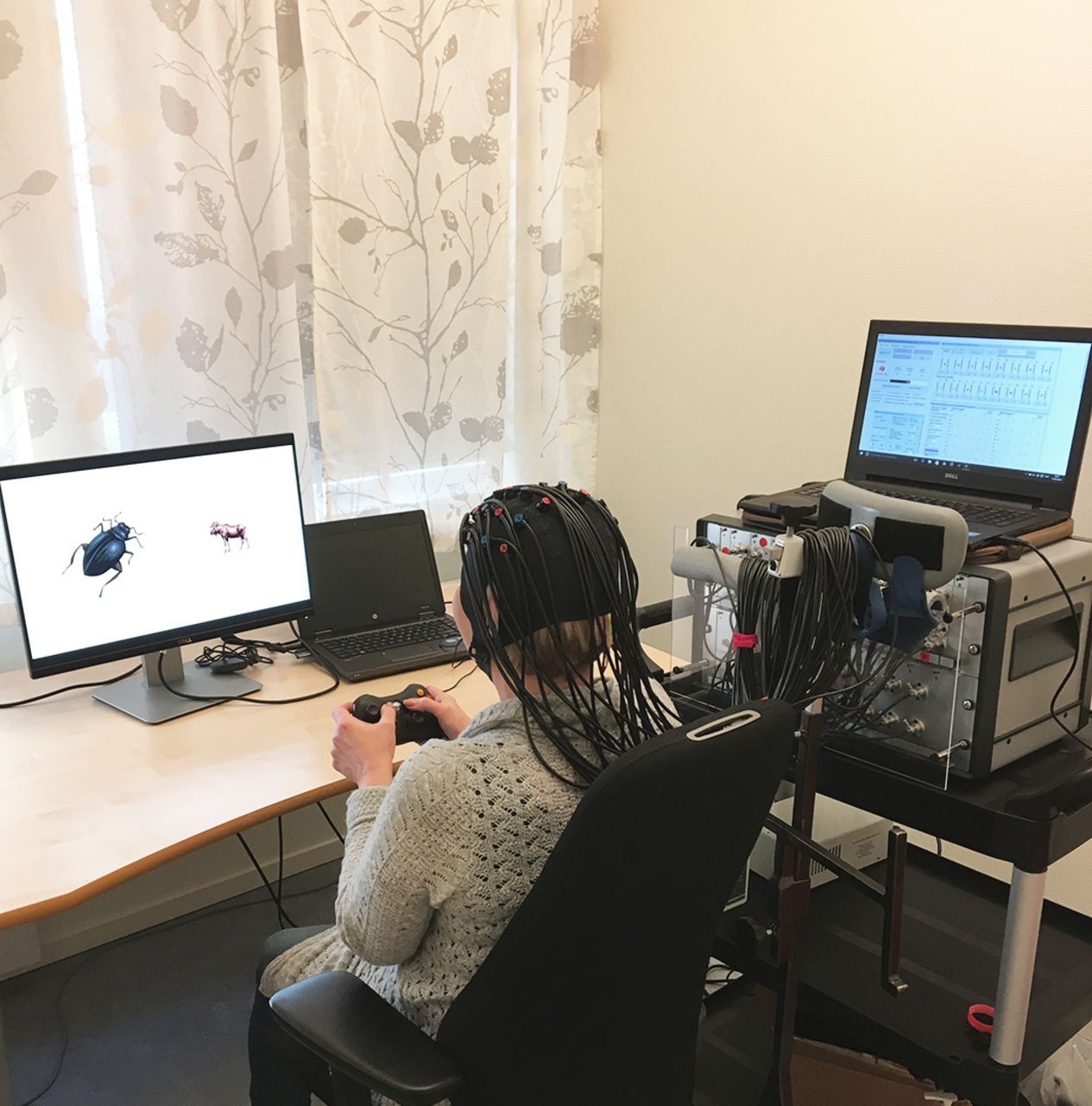
Cerebrovascular changes are measured during repeated administration of the animal Stroop test, which is modelled after the test employed by Wright et al. 80 Changes in cerebral blood oxygenation are measured by a continuous wave fNIRS device (NTS Optical Imaging System, Gowerlabs, London, UK).81 fNIRS is being increasingly used to elucidate brain function abnormalities in patients with neurological and psychiatric disorders,80 82 and non-invasively measures the concentration changes of oxygenated, deoxygenated and total haemoglobin in the cerebral cortex. The technique is based on the changes in absorption of light emitted by sources on the surface of the head and measured by detectors. Near-infrared light of two wavelengths is injected at 16 points through laser diode sources placed on the subject’s forehead and the diffused light is measured at 16 co-located detectors (figure 2). Images of animals are shown to the subject every 12 s on the left and right sides of a computer screen. The participant presses the right or left button to indicate which animal is the largest in reality, even though it may appear smaller on the computer screen. The fNIRS assessment is repeated after 30 min of testing the patient’s reading comprehension. This approach specifically taxes the tenacity that is deficient in many of these patients, enabling assessment of the neurophysiological correlates of their mental fatigability.

A fNIRS) cap. Photo of a person wearing an fNIRS cap with laser diode sources and detectors. fNIRS, functional near-infrared spectroscopy.
Time frame for the study actions
This study started in September 2012. fNIRS was added as an outcome measure in May 2015. MR hardware and software were upgraded in April 2014. To assess possible bias resulting from this change, an American College of Radiology MRI phantom83 was scanned just before the MRI upgrade and again in August 2017. The resulting phantom images were compared with quantify differences in geometric distortion and intensity mapping. Other possible differences due to the scanner upgrade will be evaluated by comparing the MAPER data from the baseline scans before the upgrade with those from the scans taken after upgrade. The last patient was included in March 2018 and the final evaluation will take place in June 2019. The longitudinal variability of the first 30 controls will be evaluated during September 2019 and the last control subject assessment is planned for October 2019.
Considerations of study design
This study focuses on the potential involvement of brain structure and function in hyperthyroidism and euthyroidism. To assess eye involvement, MR imaging of the orbits and TAO evaluation are performed in parallel and at an additional appointment between the neuroimaging assessments. Moderately severe and severe TAO impair QoL considerably84 and intravenous or oral glucocorticoid treatment in TAO management may affect brain structure volume and cognitive performance, as in Cushing’s disease.85 86 Patients on glucocorticoid treatment at baseline are therefore excluded. The orbital MRI and TAO assessments will make it possible to isolate TAO as a factor if it occurs during follow-up. More importantly, the orbital measurements may provide a biomarker (among others) for prediction of both TAO and failed cognitive recovery.
Psychiatric diseases are excluded only if there is an ongoing or recent psychosis that might interfere with full compliance and/or informed consent to participate. However, previous and current psychiatric diseases are carefully explored as psychiatric diseases are more prevalent than in a normal population, even before the onset of GD,87 and a common cause cannot be excluded. In addition, psychiatric symptoms often worsen during the phase of hyperthyroidism.88 Finally, it is important to assess the relevance of pre-existing psychiatric morbidity for cognitive prognosis.
Recovery from hyperthyroidism may take several months and many of those affected by GD still have impaired QoL after 1 year of treatment.11 As most patients are treated with ATD for 12–18 months, patients are re-evaluated after 15 months, but not later, to avoid possible effects of any recurrent GD disease. Moreover, although the CogThy study protocol accepts all treatments, only one patient had radioactive iodine treatment. This will simplify the interpretation of the results, as radioactive iodine treatment is associated with a worse QoL outcome and this could interfere with the study outcome.89
One inclusion criterion in the CogThy study is marked hyperthyroidism at the first visit, with fT4 ≥50 pmol/L or T3 ≥6.0 nmol/L. These rather high thyroid hormone levels were chosen to ensure that the patient is still hyperthyroid at the time of study inclusion. For ethical reasons, ATD treatment in a hyperthyroid patient cannot be delayed, and the inclusion and first study assessments always follow a few days after the clinical visit. These high thyroid hormone levels also make it more likely that the patient really has GD, although inclusion also requires a positive TRAb or diffuse uptake on scintigraphy.
Considerations regarding structural neuroimaging
Previous attempts to visualise and understand thyroid–brain interaction combined neuroimaging with neurophysiological and neuropsychiatric investigations.22 90 Cognition is complex and, as such, involves widely distributed functional systems and several brain regions, among which the MTL and the prefrontal cortex are generally recognised as being of central importance. The hippocampus is, however, not the only MTL structure involved in learning and memory. A useful concept is ‘the extended hippocampal system’,91 a recent improvement of the older concept ‘the limbic system’. This system, which works as a whole and subserves both memory and emotion, includes most of the MTL and also parts of the thalamus, the cingulate gyrus and parts of the frontal cortex. Therefore, there may be reversible MTL changes in other regions than the hippocampus in GD patients.
Manual volumetry is still considered the gold standard in MTL volumetry, but its inter-rater and intra-rater reliability is not optimal. Therefore, only one experienced rater will make all the definitive measurements in a single batch when all MR images are available. This rater will do a number of re-segmentations for assessing intra-rater validity. The advantages of the automatic methods, Freesurfer and MAPER, are that several other brain areas can be measured at the same time as the MTL structures are assessed and the high test–retest reliability makes it easy to combine results from analyses performed at different times.
The posterior cerebellum, among other regions, is probably relevant for cognition in GD. Experimentally induced hyperthyroidism increases brain perfusion in posterior cerebellar regions connected with cerebral networks thought to be associated with cognitive control.90 In the German version of the Auditory Verbal Learning scale, these perfusion changes are positively correlated with changes in performance.90 However, these ideas and other well-founded recent suggestions, for example, the cingulate gyrus is relevant for cognition,91 will not be specifically tested in the CogThy study.
Considerations regarding functional neuroimaging
Functional MRI22 92 and positron emission tomography studies17 18 demonstrate functional changes in neural networks in patients with hyperthyroidism compared with healthy controls. fNIRS and functional MRI probe the same physical tissue properties and give highly correlated results in cognitive tasks.93 Hitherto, fNIRS has had its main application in studies of the developing brain but has also been used in adults.94 Brain structures lying within a few centimetres from the skull can be assessed with fNIRS. This means many of the relevant frontal regions generally considered important for cognition can be assessed and many specific theories about their function in executive control, anticipation and/or memory have been proposed.95 However, Carlén96 has issued a note of caution, arguing that the often unclear definition of the ‘prefrontal’ cortex ‘… warrants a renewed focus on what the prefrontal area is and does.’96 This provides extra motivation for the fNIRS study, as the sensors with fNIRS are arranged in a helmet so that the patient can perform many activities during the testing, which is not possible with MRI. The ability to undertake advanced psychological testing during fNIRS is an advantage that is exploited in this study.
Considerations regarding the neuropsychological evaluation
MFS is the main neuropsychological assessment tool used in the study. It is a scale developed to capture mental fatigue, also described as astheno-emotional disorder,97 that is prevalent after acquired brain injury regardless of the cause.27 MFS has been validated for traumatic brain injury and stroke patients98 and is not affected by gender, education and age in patients between 18 and 65 years. The questions included in the scale have adequate internal consistency, with Cronbach’s alpha at 0.944. A cutoff at 10.5 is suggested, with higher values indicating more severe symptoms.98 In this study, MFS is used to assess mental fatigue at 0 and 15 months, with other standard neuropsychological tests being used as a complement to MFS.
Considerations regarding mechanistic factors
Apart from thyroid hormones, TRAb and other autoimmune factors, stress hormones secreted in connection with hyperthyroidism may affect the brain. Adrenal hyperactivity is noted in hyperthyroidism, but the metabolic clearance of cortisol increases99 and cortisol-binding globulins are at a low level.100 The net effect on the brain is unclear. Moreover, physiological responses to catecholamines are enhanced in hyperthyroidism,4 although the adrenergic effects from increased thyroid hormone levels on the brain have been insufficiently examined.
The possible parallel effects from glucocorticoids and the adrenergic system are difficult to disentangle within the present study, but information on treatment with beta-adrenergic blockers is collected and will be added to the model of confounding factors. In order to minimise interference from other hormonal confounders, only premenopausal women were included, as oestrogens also influence brain function.101
Thyroid hormone levels are very high in the GD patients participating in the study, although some reduction will have occurred at inclusion following the start of ATD therapy. It has been proposed that thyroid hormones may act optimally at euthyroid levels, as hyperthyroidism does not influence hippocampal neurogenesis in adult rats.51 Hippocampal metabolism may be affected by hyperthyroidism, as glucose metabolism in the limbic system is reduced17 18 and is negatively correlated with both serum fT3 and fT4 levels in hyperthyroid patients.18 ATD treatment corrects this hypometabolism.17
D2 activity is thought to be reduced in hyperthyroidism in order to protect tissues from thyroid hormone overexposure.37 However, this mechanism may be different in GD. Sera from GD patients with high TRAb stimulate D2 activity,49 102 which partially explains why GD patients have more severe mental symptoms than patients with other forms of hyperthyroidism.13
The problem of long-term cognitive dysfunction has been addressed in a study on twins concordant or discordant for earlier hyperthyroidism. Although there were no differences, the authors noted that the study may have been underpowered due to the small number of twin pairs discordant for hyperthyroidism.103 Finally, it has been suggested that hyperthyroidism accelerates the ageing process,30 but this hypothesis has not been evaluated.
Power calculation and statistical considerations
Changes over time and differences between groups concerning volumetric and functional imaging data will primarily be analysed with univariate statistical methods. Due to a lack of previous studies on this topic when the study was conceived, the power calculation for the volumetric analysis was based on changes in hippocampus volume in patients with Cushing’s disease scanned with a 1.5 T scanner. A sample of approximately 40 subjects was considered sufficient for within-individual comparison to detect a mean volumetric change of 10% with an 80% probability and 5% significance. With a 3 T scanner, smaller changes can be reliably detected. Allowing for expected attrition, at least 60 patients need to be included to have sufficient power. We will follow the dropout frequency regularly and adjust sample size to attain sufficient power. These numbers relate to paired comparisons of successive manual segmentations of the hippocampus. It is generally agreed that longitudinal comparisons with serial registration methods are more sensitive and such methods may be included. Despite the lack of published longitudinal MRI studies of MTL structures in thyroid disease, we are confident that the study power will be sufficient to detect any clinically meaningful changes in hippocampal volume over time.
For the fNIRS power calculation, data from an ongoing fNIRS study at Sahlgrenska University Hospital, Göteborg on a similar patient group (traumatic brain injury) and with the same study design as the current one have been used. It was assumed that the general trend of the blood flow changes would be similar in the two studies. Based on an unpaired one-sided t-test with a significance level of 0.05 and a power of 0.8, the required sample size for detecting the same difference between groups, as in the traumatic brain injury study, is 21 patients and 26 controls. With equal distribution, 23 subjects are needed in each group. The controls and patients are matched according to age, sex, smoking and educational level. A paired t-test will be used and the power of the paired test is expected to be higher than that of the unpaired test (and it cannot be lower), in which case the sample size needed will be smaller.
The associations between the volumetric data and the other study variables such as MFS score, QoL, laboratory data and psychiatric assessments will be analysed with both univariate and multivariate statistics. Principal component analysis and similar methods will be used to trace common causal factors. No independent power calculations have been performed for these analyses.
The research group has a general aim to suggest a causal and predictive model for mental fatigue in GD. The formulation of this model will take background knowledge from neuropsychiatry, neuroimaging, neuroendocrinology and neuroimmunology as its point of departure. Data from the present project, a planned continuation of it and published data will be used as inputs. The final model will be framed as a hypothesis to be validated in further studies.
Summary
Impaired well-being in GD patients is a long-term problem and is frustrating for both patients and healthcare. It is not understood why this happens and it cannot be predicted at an early stage which patients will develop long-term cognitive impairment. Hence, the CogThy project addresses a considerable clinical problem. This project will: (1) identify morphological brain features associated with psychiatric outcome and thyroid hormone function in GD patients and (2) use fNIRS to study cognition in GD patients. Planned future achievements of this project are: (1) to develop a risk factor model that will weight all possible factors for identifying subjects with the highest risk of developing persistent cognitive impairment and (2) the identification of biomarkers for cognitive deterioration that can be used for risk evaluation and monitoring
References
Footnotes
Contributors MOH has written the manuscript with input from all the other authors. HM is responsible for the statistics and has broad knowledge about mental syndromes. As a psychiatrist, PB will perform psychiatric evaluations and, as neuropsychologist, BJ will perform psychological testing. LB-K and SS will perform and evaluate fNIRS investigations, and EO, RAH and NK are responsible for volumetry evaluations. MOH is a PhD student in the project and will see all patients in collaboration with HNF, who is the principal investigator of the CogThy project.
Funding The following sponsors supported this study. The study was financed by grants from: the Swedish state under an agreement between the Swedish government and county councils, the ALF agreement (ALFGBG-717311); regional research funding, Region Västra Götaland; the Healthcare Sub-committee, Region Västra Götaland; the Healthcare Board, Region Västra Götaland; Sahlgrenska University Hospital research funds; the Gothenburg Medical Society; the Swedish Medical Society; the Swedish Society for Medical Research; The Swedish Endocrine Society; The Fredrik and Ingrid Thuring's Foundation; the Iris grant; the Jeanssons's Foundation; the Tore Nilsson's Foundation; the Wilhelm and Martina Lundgren's Foundation; the Pharmacist Hedberg's Foundation and the Åke Wiberg's Foundation. Siemens Healthineers has supported the study by providing reagents for TSI. The project is also supported by MedTech West ( www.medtechwest.se ), a medical innovation and development platform initiated as a collaboration between Chalmers University of Technology, University of Borås, University of Gothenburg, Sahlgrenska University Hospital and Region Västra Götaland, Sweden. MedTech West provides office space for the project participants as well as laboratory space and meeting facilities for activities of the CogThy project. The supporting or funding bodies have no role in the study design, collection, analysis and interpretation of data, in manuscript writing or in the decision to submit the manuscript for publication.
Competing interests HNF has received lecture fees from Siemens Inc., AstraZeneca and Bristol-Myers Squibb.
Patient consent for publication Not required.
Ethics approval Ethical approval was granted by the Regional Ethical Review Board (Ref no. 190-10; approved 21 June 2010) in Göteborg, Sweden. The study is conducted according to the Declaration of Helsinki. The results will be actively disseminated through peer-reviewed journals, national and international conference presentations and among patient organisations after an appropriate embargo time. Preliminary data have been presented at: the Brain's Networks conference in Gothenburg 2015; the Annual Meeting of the European Thyroid Association in Copenhagen 2016; the Annual Meeting of the American Thyroid Association in Victoria, Canada 2017; the Annual Conference of the Swedish Endocrine Society 2017 and the American Thyroid Association Annual Meeting in Washington, USA, 2018.
Provenance and peer review Not commissioned; externally peer reviewed.