Article Text

Abstract
Introduction 2-D ultrasound shear wave elastography (SWE) could be considered as a new noninvasive tool for monitoring fetal lung development based on evaluation of mechanical properties during pregnancy. Interesting results are available concerning the use of SWE on developing organs, especially on premature infants and animal models. The main objective in this study is to evaluate the feasibility of 2-D SWE in human fetal lungs between 24 and 34 weeks of gestation (WG). The secondary objective is to modellise fetal lung-to-liver elastography ratio (LLE ratio) and to assess variations between normal lung and lung surfactant-enriched after a corticosteroids course indicated for a threatened preterm labour (TPL).
Methods/design A prospective case-control study will be performed between 24 and 34 WG. Fetal lungs and liver will be explored by SWE into two groups: fetuses of women with an uncomplicated pregnancy (control group) and fetuses of women with a TPL requiring administration of corticosteroids (cases group). LLE ratio will be defined as the value of the lung elasticity divided by the value of the liver elasticity.
Primary judgement criterion is the value of elasticity modulus expressed in kilopascal. Lungs and liver will be explored through three measurements to define the most reproducible regions with the lowest intra- and inter-observer variability. Feasibility will be evaluated by assessing the number of examinations performed and the number of examinations with interpretable results. Intra- and inter-observer reproducibility will be evaluated by means of the intra-class correlation coefficient.
Ethics and dissemination Approval of the study protocol was obtained from the human ethical research committee (Comité de Protection des Personnes EST II, process number 15/494) and the French National Agency for Medicines and Health Products Safety (process number 2015-A01575-44). All participants will sign a statement of informed consent.
Trial registration number NCT02870608; Recruiting.
- Shear wave elastography
- elasticity
- fetal
- lung.
This is an Open Access article distributed in accordance with the Creative Commons Attribution Non Commercial (CC BY-NC 4.0) license, which permits others to distribute, remix, adapt, build upon this work non-commercially, and license their derivative works on different terms, provided the original work is properly cited and the use is non-commercial. See: http://creativecommons.org/licenses/by-nc/4.0/
Statistics from Altmetric.com
Strengths and limitation of the study
This study will be the first one to demonstrate applicability of shear wave elastography in human fetal lungs.
Pharmacovigilance data will be collected at birth and 3 months later in order to assess safety of the device.
Different factors will be taken into account to understand variability of measures: acquisition depth, number of measurements and representative values such as median or mean values.
We shall not attempt to define reference value of fetal lung elasticity or to correlate SWE value with respiratory function at birth.
Introduction
Background
Prenatal evaluation of fetal lung maturity (FLM) is still a challenge and different approaches have been reported with invasive biological tools or fetal imaging including ultrasounds and MRI. FLM is mainly correlated with surfactant level and numerous tests have been developed to quantify this production: lecithin/sphingomyelin ratio (L/S), laminar body count, testing for Phosphatidyl glycerol with thin-layer chromatography and estimation of the surfactant/albumin ratio using polarised light. All of these tests have been shown to have good sensitivity and negative predictive values. However, they all have a low positive predictive value and need invasive procedure.1 2 Thus, amniocentesis for FLM became obsolete.3
Lung and liver signal or echogenicity were often compared as ratio to assess FLM in several studies.4–10 Non-invasive assessment of FLM has been first attempted using ultrasonography quantified by grey level histogram.4 11 12 These studies demonstrated sonographic changes associated with lung maturation but weak correlations between echogenicity and respiratory distress syndrome (RDS). Quantitative analysis with specific software has been secondly proposed to improve robustness. Fetal lung texture analysis with this method demonstrated a strong correlation with gestational age, biological test on amniotic fluid and risk of neonatal respiratory morbidity.13 14 Additionally, several studies found a significant association between fetal lung-to-liver signal intensity ratio and estimated gestational age with fetal MRI. Actually, this technique is mainly used to assess fetal lung development and volume, especially in case of congenital diaphragmatic hernia or severe oligohydramnios. Nevertheless, the accuracy of both MRI and ultrasounds for estimating long-term respiratory outcomes remains limited.15–19 These previous techniques propose prenatal evaluation of lung maturity based on imaging features, but prediction of the pulmonary function through prenatal biomechanical properties seems relevant. With this hypothesis, we advanced the concept of ‘prenatal functional imaging’ and shear wave elastography (SWE) could be a promising tool.
SWE is a recent ultrasound technology using acoustic radiation force imaging (ARFI), which enables the assessment of the stiffness of tissues in real time through a colour quantitative elastogram.20 SWE assesses tissue elasticity (E), which is the tendency of tissue to resist deformation with an applied force. In a locally homogeneous and purely elastic medium, we can calculate Young’s modulus (kPa) from the shear wave speed (m.s-1) with the formula: E=3ρ.csw² (ρ being the medium density and csw the shear wave speed). The stiffness at any location of the region of interest (ROI) can be sampled using measurement tools to obtain a quantitative evaluation either in terms of shear wave speed or kPa. A low shear wave speed corresponds to a soft tissue, while a shear wave high speed indicates a stiff tissue. The study of deep organs is possible with SWE because this method does not require any compression-relaxation sequence on the target organ. One of the advantages of SWE over other elastography methods is that the generation of the mechanical impulse is operator-independent. SWE is expanding its range nowadays by its promising role in the examination of various organs (liver, thyroid) and for aiding discrimination of lesion characteristic, especially breast or prostate tumours.21–24
There is to date no publication regarding its use on human fetuses. The Food and Drug Administration (FDA) has so far not yet approved the use of SWE for obstetric clinical applications because of the paucity of data in the literature. Although there is no report about apparent histological changes with shear wave, the absence of other bioeffects could not be discarded and further studies are recommended.25 However, setting parameters respect mechanical and thermal indexes for soft tissues and bone, which are necessary to allow an obstetric examination defined by the FDA.26 Thus, there is a debate concerning the safety of SWE in fetal medicine. Herman et al studied the models and regulatory considerations for transient temperature rise during ARFI, and found that any transient increase in temperature caused by pulse bursts might still be within the safe limits determined by the FDA.27 Other authors showed that transducer heating was below 1°C for the current clinical applications of ARFI. According to experimental studies that simulate the heating of soft tissue during ARFI, they demonstrate that ARFI on soft tissue is safe, provided that thermal index be monitored.28 29
Interesting and reassuring results are available concerning the use of SWE on developing organs, especially on premature infants and animal models. Principal organs explored are the liver, brain and lungs. Quarello et al demonstrated the possibility of using SWE in non-human primate fetuses and its feasibility in exploring fetal organs. They found that elasticity values were related to organs and gestational age.30 Concerning fetal lungs elasticity, measurements of the proximal lungs seem to increase throughout the pregnancy. Concerning the use of SWE on premature infants, results are more numerous and include exploration of the liver or the brain. Alison et al have demonstrated the feasibility and reproducibility of liver stiffness in preterm neonates with intra uterine growth restriction (IUGR) and gestational age at birth between 26 and 31 weeks of gestation (WG).31 Other studies reported the contribution of SWE to the diagnosis of biliary atresia in neonates.32 33 Kim et al described the variation of brain elasticity in different regions in healthy neonates born between 28 and 40 WG.34 Su et al quantitatively evaluated the effect of ARFI in neonatal brain development. The authors found that full-term neonates had significantly higher elasticity values than preterm neonates and there were no reported immediate adverse events.35 Other teams work on the application of this technology to neonates as a supplementary tool to detect early ischaemic brain injury.36
If we describe fetal thorax centred on a four-chamber view of the fetal heart with SWE, the following observations can be made (figure 1)

(A) B-mode image of the fetal thorax centred on a four-chamber view of the fetal heart, using an abdominal convex probe 1–6 MHz. (B) Elastogram of the fetal thorax showing a colour-coded elasticity map: blue identifies deformable tissue and red indicates rigid tissues (kPa).
SWE is able to make distinction between fetal soft and stiff tissues. Fetal ribs appear red coloured mapped, whereas proximal lung appears blue coloured.
A homogeneous coloured area can be obtained on the proximal lung with a colour scale ranging from 0 to 48 kPa. A homogeneous ROI is important for the reliability of the results.
SWE can differ according to the acquisition depth. Proximal lung appears with a blue homogenous distribution whereas distal lung appears with green homogenous distribution.
Objectives
The main objective in this study is to evaluate the feasibility of 2-D SWE in human fetal lungs between 24 and 34 WG. The secondary objective is to modellise fetal lung-to-liver elastography ratio (LLE ratio) and to assess variations between normal lung and lung surfactant-enriched after a corticosteroids course indicated for a threatened preterm labour (TPL).
Methods/design
Study design and setting
A prospective case-control study will be performed at the University Hospital of Besançon, (Besançon, France), Department of Obstetrics and Gynaecology. Fetal lungs and liver will be explored by SWE between 24 and 34 WG in two groups: fetuses of women with an uncomplicated pregnancy will be considered as the control group and fetuses of women with a threatened preterm labour requiring administration of corticosteroids will be enrolled as cases. The first eligible patient was enrolled in May 2016 and we plan to enrol for 18 months. End of follow-up of children will be completed approximately 6 months after the last birth.
In this pilot study, corticosteroids generate an effect model interesting for the assessment of LLE ratio variation in the cases. We take advantage of the action of corticosteroids to explore another medium with different supposed biomechanical properties. Indeed, corticosteroids accelerated the development of type two pneumocytes leading to surfactant production. Similarly, there is a significant increase in saturated phosphatidylcholine content in the fetal lung, potentially modifying the propagation of shear waves.37
The control group will be recruited during routine prenatal visits or ultrasounds according to the French ultrasounds schedule (ultrasound screening during the second trimester between 20 and 25 WG and the third trimester between 30 and 35 WG).38 Cases will be enrolled during their hospitalisation for threatened preterm delivery (figure 2). The design, conduction and reporting of this study will follow the norms of the Strengthening the Reporting of Observational Studies in Epidemiology (STROBE) statement.39
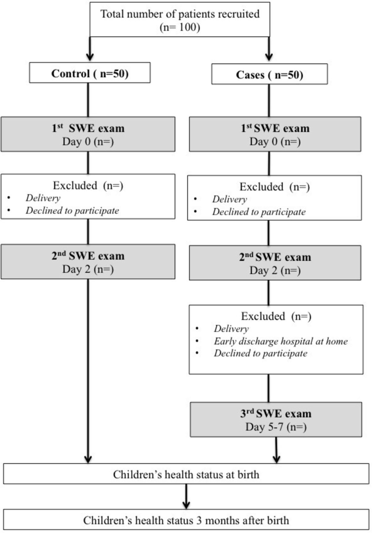
Design of the study.
Ethical considerations
The study respects the ethical standards established in the Declaration of Helsinki.40 Investigators undertake to respect law no. 2004–806 of 9 August 2004 relative to public health policy on clinical trials, made effective by implementing Decree 2006–477 of 26 April 2006.41 Approval of the study protocol was obtained from the human ethical research committee (Comité de Protection des Personnes EST II, process number 15/494) and the French National Agency for Medicines and Health Products Safety (process number 2015-A01575-44). The study is registered on clinicaltrial.gov with the following number: NCT02870608. All participants will sign a statement of informed consent and will be allowed to abandon the study at any time, without negative consequences. All procedures of the study will be confidential. The study will involve the collection and storage of ultrasound images, and study participants will therefore be thoroughly informed. All variables (patient parameters, data items, data elements) will be aggregated into electronic case report forms.
As explained in the background, the FDA has so far not yet approved the use of SWE for obstetric clinical applications because of the paucity of data in the literature. In this study, fetuses will be included from 24 to 34 WG, over the period of organogenesis. In the patient information sheet, it is specified that previous studies explored the liver or brain of premature infants with SWE. Alison et al studied liver stiffness in preterm neonates with IUGR and gestational age at birth between 26 and 31 WG.31 Franchi-Abella et al evaluate the feasibility and diagnostic accuracy of SWE for the assessment of liver stiffness in 96 patients, and seven were preterm newborns between 30 and 35 WG.42 Both studies did not report adverse outcomes and were approved by an institutional review board. Moreover, the consent form mentions conclusions of an experimental study of the biological effects associated with SWE in brains of neonatal mice. Results indicated that using SWE does not cause detectable histological changes. Potential effects on intracellular signalling pathways were detected when the SWE scanning lasted more than 30 min.25 Thus, patients will be informed that our study is focused on fetuses with a gestational age similar to preterm newborn included in the above-mentioned studies. Moreover, exploration with SWE will concern only lung and liver with duration of less than 10 min in respect of biophysical safety indexes: Thermal index Ti≤0.7 and Mechanical index Mi<1,9.
Participants
Cases will be recruited during hospitalisation for a threatened preterm labour requiring the administration of corticosteroids. The complementary inclusion criteria are: pregnant women aged 18 years or more; singleton pregnancy with eutrophic fetus; signature of consent; and affiliation to health insurance scheme (figure 2). Patients will be stratified by gestational age between 24 and 34 WG, with one class every WG (10 classes in total) and five patients per class. After obtaining consent, a SWE examination will be performed before the first intramuscular administration of corticosteroids ‘day 0’ (betamethasone 12 mg). A second SWE examination will be performed within 24 hours after the second intramuscular administration of corticosteroids ‘day 2’. A third SWE will be performed between the fifth and the seventh day following the first administration of corticosteroids ‘day 5–7’, if the women are not early discharged at home.
For the control group, women will be matched by gestational age and will be recruited during their pregnancy monitoring. Information concerning the study will be given as early as the second trimester by obstetricians or midwives during medical visits or ultrasounds. The complementary inclusion criteria are the following: major pregnant women; uncomplicated pregnancy between 24 and 34 WG; singleton pregnancy with eutrophic fetus; signature of consent; and affiliation to health insurance scheme. If they are willing to participate, a first SWE examination will be proposed at ‘day 0’, followed by a second one in 2 days ‘day 2’. Patients who come back for an SWE examination performed outside the current medical visit will receive financial compensation for travel expenses. As well as cases, control patients will be stratified by gestational age between 24 and 34 WG.
Exclusion criteria common to both groups are: fetal lung or liver pathologies; inclusion in another medical study; and patients under legal incapacity.
Variables and measuring technique
Prenatal variables
Primary judgement criterion is the value of kPa (E) (elasticity modulus) expressed in kilopascal (kPa). A Logic E9 system (General Electric Medical System, Milwaukee, WI, USA) with CE certification CE LNE/G-MED [CE0459] will be used for this study, equipped with abdominal convex probe 1–6 MHz (C1-6-D probe). This technique includes a real-time visualisation of a colour quantitative elastogram coupled to a B-mode image. To provide robust shear wave and optimise its detection, Logiq E9 uses innovative techniques. Comb-push ultrasound SWE generates multiple shear wave sources into theROI and uses directional filtering to remove the interference. The TAST technique is able to robustly track shear waves and correct for the sequential tracking delay.43 This technique has the merit of rapid and solid reconstruction of a large elasticity map (elastogram) with the only single acquisition by generating multiple shear waves from multiple unfocused push beams.
The shear wave acquisition measurement protocol is saved in a specific preset that will be used for every patient: 2D-Mode: harmonic imaging central depth=6 cm, frequency=6 MHz, acoustic power 100%; Elastography-Mode: shear wave signal frequency ranges from 50 to 400 kHz, acoustic power of the pushing beam: 100%, Tracking power: 100%, colour scale ranging from 0 to 48 kPa. Biophysical safety indices: Thermal index Ti≤0.7 and Mechanical index Mi<1,9 (table 1).
Fetal lungs will be voluntarily approached laterally to systematically obtain proximal and distal lungs regarding the distance to the probe. Each measurement has to be performed on a homogeneous elastogram to be valid. Operators will perform two cycles of nine kPa measurements, while systematically repositioning the probe on each target organ.
A cycle includes: three measurements on the proximal lung (anterior ‘P1’, medium ‘P2’ and posterior portion ‘P3’); three measurements on the distal lung (anterior ‘D1’, medium ‘D2’ and posterior portion ‘D3’); and three measurements on three liver segments (IV, V, VI) (figure 3). Implementation of SWE displays 2D images of kPa in the target organ. In real time, the elasticity appears colour-coded, where blue identifies deformable tissue and red indicates rigid tissues. The kPa value at any location will be sampled using a round ROI of 5 mm. The distance between the probe and the target organ will be collected. The average elasticity in the ROI will be automatically recorded by the system in a worksheet. The investigators will adjust the position of the ROI using the B-mode image for guidance and take care to obtain the most homogeneous colour-coded ROI before kPa estimation (figure 4). Technical failure was defined as failure to obtain a homogeneous elastogram in more than 50% in the sampling area. To test the inter-observer variability, a second observer will successively perform measurements on 30 fetuses. All measurements will be carried out directly by ultrasound on the target organ. The second observer will perform measurements just after the first one and will be blinded to the feasibility and results obtained by the first one.

Measurement sites with 2D comb-push SWE using an abdominal convex probe 1–6 MHz (C1-6-D probe). Colour scale ranging from 0 to 48 kPa. Sufficient colour maps covering more than 50% of the sampling area obtained considered as a technical success. ROI are placed on homogeneous elastograms.- (A,B) three measurements on the proximal lung (anterior ‘P1’, medium ‘P2’ and posterior portion ‘P3’). - (A,C) three measurements on the distal lung (anterior ‘D1’, medium ‘D2’ and posterior portion ‘D3’). - (D–F) three measurements on three liver segments (IV, (V, VI). Operator will perform two cycles of nine kPa measurements while systematically repositioning the probe and each target ROI.

2D comb-push SWE on proximal fetal lung. Colour scale ranging from 0 to 48 kPa. On image (A), elastogram is not homogeneous and is degraded by artefact on the left side due to rib shadowing. By moving the probe to another location, a more homogeneous elastogram (B) is obtained allowing measurement.
Estimation of Fetal Weight Estimation (EFW) will be performed during each examination according to the Hadlock formula based on cephalic circumference (CC), abdominal circumference (AC) and femoral length (FL): log10 EFW=1326 + 0,0107 PC+0,0438 PA+0158 LF+0 00 326 (PA x LF).44
Postnatal variables
The following pharmacovigilance data will be collected at birth and 3 months' later in order to assess safety of the device:
At birth in both groups: term of birth, weight, neonatal transfer, Apgar Score and evaluation of respiratory distress by Silverman Score.45
Three months after birth: medical history, health problems, respiratory diseases or symptoms, liver disease or symptoms and number of hospitalisations since birth.
Limitation of bias
The use of sampling method with five patients per class of gestational age will limit repartition bias between control and cases. Inclusion of any pregnant women attending Besançon University maternity department for the control group, and inclusion of all women with a threatened preterm labour will limit the risk of selection bias. Risk of loss will be limited in the cases group between ‘day 0’ and ‘day 2’ because patients will remain hospitalised for corticosteroids administration. Nevertheless, the risk of loss is most important between ‘day 2’ and ‘day 5–7’ because of possible early discharge at home after the second administration of corticosteroids. For ethical and medical reasons, cases early discharge at home cannot be convened in ‘day 5–7’. Risk of loss will be limited in the control group because of a financial compensation for travel expenses. To limit inter-observer variation, expert sonographers will perform elastography examinations.
Study size
Because of the paucity of data regarding SWE on the human fetal lung, the sample size calculation was based on measurements of fetal lungs in pregnant baboons and extrapolations made by clinicians, with the following assumptions: average expected value of elasticity coefficient for fetal lung will be 2 kPa at 24 WG and 4 kPa at 34 WG. We hypothesised a linear increase of elasticity coefficient during pregnancy in the control group and a decrease in cases exposed to corticosteroids. At ‘day 2’, we therefore expect in the cases a variation of 0.2 kPa (corresponding to 1 week of lung maturation during gestation). Forty-two patients per group will be needed to have 90% power to statistically demonstrate such a difference (0.2 vs 0.057) assuming α risk of 0.05 and SD of 0.2.
Proposed statistical analysis
Technical validation
Feasibility will be evaluated by assessing the number of examinations performed and the number of examinations with interpretable results. All the variables and data that will be stored for each SWE examination are summarised in table 1. Intra- and inter-observer reproducibility will be evaluated by the intra-class correlation coefficient (ICC) with 95% CI. Intra-observer reproducibility will be calculated using the two cycles of measure, inter observer reproducibility between the operators will be calculated by means of the two cycles of measures if the intra-observer is high.46 Only repeatable and reproducible values of each ROI will be considered to modellise the LLE ratio.
Variables and data stored for each SWE examination. ROI (region of interest)
Clinical evaluation
Evolution of fetal lung SWE, fetal liver SWE and lung-to-liver-SWE ratio will be assessed by group and overall. Lung-to-liver-SWE ratio will be initially defined as the value of the lung elasticity divided by the value of the liver elasticity.
SWE values before corticosteroids administration will be compared between cases and control groups. If there is a statistically significant difference, a confusional bias will have to be sought between fetal lung elasticity values and threatened preterm labour. All results will be presented as ‘delta’ variation between two measurements in order to limit ‘non-comparability bias’ between both groups. If corticosteroids affect fetal lung elasticity, ‘delta before/after’ will be more important in cases than controls.
Analysis of mean differences between groups will be carried out using a Student’s test or Wilcoxon test according to the distribution of data for quantitative variables. The Shapiro–Wilk test will be used to determine if the data set is well modelled by a normal distribution. The relationship of quantitative variables to each other will be tested using Pearson’s or Spearman’s correlation as appropriate.
Qualitative variables will be expressed as frequencies and quantitative variables will be displayed as the mean ±SD if normally distributed or as the median ±IQR if asymmetrically distributed. Qualitative variables will be analysed using a χ2 test or Fisher test.
For statistical analyses, the level of statistical significance will be set at 5% (P<0.05). Statistical analysis will be performed with statistical software SAS for Windows, version 9.4 and MedCalc software, version 15.
Discussion
2D-SWE could be considered as a new noninvasive tool for monitoring fetal lung development based on the evaluation of mechanical properties during pregnancy. However, standardisation for measuring SWE in the human fetal lung is necessary to demonstrate applicability of the technique, and to understand limiting factors. This pilot study will focus on technical performances before considering clinical applications. In this protocol, SWE will be used after the fetal period of organogenesis, and pharmacovigilance data will be collected at birth and 3 months' later in order to assess the safety of the device.
With the study design, we shall not attempt to define the reference value of fetal lung elasticity during pregnancy or to correlate SWE value with respiratory function at birth. The main objective is to evaluate the feasibility and reproducibility of 2D-SWE in the human fetal lung between 24 and 34 WG and to modellise a new LLE ratio. Indeed, each target organ will be explored through three measurements to define the most relevant and reproducible regions with the lowest intra- and inter-observer variability. The use of the LLE ratio is interesting because it allows comparison between two tissues in which the liver is taken as a reference organ. Several studies indicate that liver signal or echogenicity remain constant through gestation, suggestive of a regular evolution of the tissue characteristics.47 48 Thus, we will compare the evolution of SWE values between two organs subjected to the same technical variations, especially for depth. Both lung and liver tissue density increase during pregnancy and we expect a linear increase of SWE values of these organs. In case of variations of lung SWE values after a course of corticosteroids, LLE ratio should be modified. If we suppose that liver elasticity will be constant between two separate examinations of 48 hours, the LLE ratio will decrease if corticosteroids lead to attenuation of shear wave speed. Conversely, if lung elasticity increases after a course of corticosteroids, the LLE ratio will also increase. Finally, the LLE ratio will be used to increase the reliability of the results and to standardise the study.
One advantage of this protocol is to evaluate SWE in three tissues with different supposed biomechanical properties: normal lung, lung with increase of surfactant synthesis after corticosteroids course and liver. Corticosteroids generate an effect model interesting for the assessment of LLE’s ratio variation in the cases because of a significant increase of phosphatidylcholine content in fetal lungs. This effect is mediated by an activation of choline-phosphate cytidylyltransferase.37 Given the placental transfer and time to induce the cellular mechanism of transcription, an artificial delay of 24 hours seems to be necessary to produce surfactant in pneumocytes type 2. The benefit of corticosteroid administration is greatest at 2 to 7 days after the initial dose.49 50 Two approaches can be proposed after a complete course of corticosteroids. On the one hand, the SWE of the fetal lung could increase because corticosteroids accelerate FLM, and we could expect an advanced ripening by one WG. On the other hand, an increase in phospholipids content in fetal lung after corticosteroids could increase viscosity and lead to dispersion of shear wave speed and attenuation.
Different factors will be taken into account in this study to understand the variability of measures: acquisition depth, number of measurements and representative values such as median or mean values. Shear wave velocity can be different according to the acquisition depth and this parameter is gradually underestimated with increasing depth. Effect is greatest in stiff tissues but little effect is seen in a soft target.51–53 This phenomenon might be explained by a damping of the acoustic push pulse that generates the shear waves by both increased attenuation and increased stiffness of the target.54 Thus, we expected low variation between proximal and distal lung because they may be considered as soft tissues. There are also debates about the acquisition number during SWE and the designers of the device did not provided advices. The number of measurements (NMs) reported in the recent literature on various organs is inconsistent (commonly seen are 3, 4 and 5 NMs).55 56 Quarello et al reported 3 NMs on fetal lungs in pregnant baboons, and ICC for and inter-observer variability were better for an average value of three measurements.57 For Alison et al, 9 NMs were performed on liver in preterm neonates with intra uterine growth restriction (three measurements from three different liver segments). Measurements showed high reproducibility on average values (ICC=0.94–0.98 for intra-operator, 0.86 for inter-operator).31 One objective of our protocol is to determine the NMs necessary to evaluate fetal lung SWE with a good inter- and intra-observer reproducibility. Thus, each value will be analysed to determine if mean lung and liver SWE can be achieved through only one valid measurement or more, and if its performance is equivalent to two or three valid measurements.
To sum up, we hope that the results of this study will contribute to clarify the applicability of SWE on the human fetal lung between 24 and 34 WG. SWE could be a new tool for the reliable prediction of lung development through biomechanical properties. This study will be the first one to propose a protocol of measurement underlying limiting factors.
References
Footnotes
Contributors All the authors contributed to the conception and design of the study. NM, SA and RR provided the idea for the research or article, created the hypothesis and wrote the original proposal. NM, CV, LP and RR significantly contributed to writing the paper. NM, CV, GB and LP wrote this protocol paper. All authors read and approved the final manuscript.
Funding This study is supported by University Hospital of Besançon, APICHU Réf: API/2015/60.
Competing interests None declared.
Patient consent Obtained.
Ethics approval The Human Research Ethics Committee (Comité de Protection des Personnes EST II) process number 15/494.
Provenance and peer review Not commissioned; externally peer reviewed.