


Abstract
Chronic abuse of methamphetamine leads to cognitive dysfunction and high rates of relapse, paralleled by significant changes of brain dopamine and serotonin neurotransmission. Previously, we found that rats with extended access to methamphetamine self-administration displayed enhanced methamphetamine-primed reinstatement of drug-seeking and cognitive deficits relative to limited access animals. The present study investigated whether extended access to methamphetamine self-administration produced abnormalities in dopamine and serotonin systems in rat forebrain. Rats self-administered methamphetamine (0.02-mg/i.v. infusion) during daily 1-h sessions for 7 to 10 days, followed by either short- (1-h) or long-access (6-h) self-administration for 12 to 14 days. Lever responding was extinguished for 2 weeks before either reinstatement testing or rapid decapitation and tissue dissection. Tissue levels of monoamine transporters and markers of methamphetamine-induced toxicity were analyzed in several forebrain areas. Long-access methamphetamine self-administration resulted in escalation of daily drug intake (∼7 mg/kg/day) and enhanced drug-primed reinstatement compared with the short-access group. Furthermore, long-, but not short-access to self-administered methamphetamine resulted in persistent decreases in dopamine transporter (DAT) protein levels in the prefrontal cortex and dorsal striatum. In contrast, only minor alterations in the tissue levels of dopamine or its metabolites were found, and no changes in markers specific for dopamine terminals or glial cell activation were detected. Our findings suggest that persistent methamphetamine seeking is associated with region-selective changes in DAT levels without accompanying monoaminergic neurotoxicity. Greater understanding of the neuroadaptations underlying persistent methamphetamine seeking and cognitive deficits could yield targets suitable for future therapeutic interventions.
- Meth, methamphetamine
- DAT, dopamine transporter(s)
- SERT, serotonin transporter(s)
- dSTR, dorsal striatum
- NAc, nucleus accumbens
- PFC, prefrontal cortex
- HPLC, high-performance liquid chromatography
- DOPAC, 3,4-dihydroxyphenylacetic acid
- HVA, homovanillic acid
- GFAP, glial fibrillary acidic protein
- Iba, ionized calcium-binding adaptor molecule
- ANOVA, analysis of variance
- vPFC, ventral prefrontal cortex.
Methamphetamine (Meth) abuse in humans can quickly develop into a chronic relapsing disorder, accompanied by a wide range of neuropsychological deficits. For example, Meth addicts display impairments in memory functions, cognitive and psychomotor performance, as well as increased impulsivity and aggressive behavior (for reviews, see Nordahl et al., 2003; Scott et al., 2007). Human brain imaging studies provide evidence that these neuropsychological deficits are paralleled by significant changes in brain dopaminergic and serotonergic neurotransmitter systems, as well as altered general metabolic activity in basal ganglia and frontal cortices (for review, see Chang et al., 2007). In particular, chronic Meth abuse reduces the density of dopamine transporters (DAT) in the striatum and (to a lesser extent) in the frontal cortex of abstinent Meth users (Volkow et al., 2001; Sekine et al., 2003; McCann et al., 2008). Similarly, a decreased number of serotonin transporters (SERT) was detected across several brain regions in abstinent Meth addicts (Sekine et al., 2006).
Most frequently, animal models of repeated Meth exposure have used short-term noncontingent, multiple high-dose (“binge”) regimens producing Meth-induced neurotoxicity, specifically damaging dopamine and serotonin terminals in the brain. As a consequence, persistent reductions in striatal dopamine content, DAT density, and activity of the dopamine-synthesizing enzyme tyrosine hydroxylase have been described in brains of rodents and nonhuman primates (for review, see Volz et al., 2007). It has been repeatedly observed that Meth-induced insult to dopamine terminals was more severe in the dorsal striatum (dSTR) in comparison with nucleus accumbens (NAc) and prefrontal cortex (PFC), probably reflecting the different densities of DAT in these regions (Prudencio et al., 2002; Volz et al., 2007). It is well established that DAT plays a major role in cellular mechanisms of Meth-induced neurotoxicity. Thus, mice with a genetic deletion of DAT are significantly less vulnerable to the neurotoxic effects of Meth (Fumagalli et al., 1998; Numachi et al., 2007). Disruption of dopamine homeostasis by high Meth concentrations (in the striatum) leads to increased oxidative stress, activation of apoptotic cascades and inflammatory mediators, as well as the activation of microglia and astrocytes (Thomas et al., 2004; Quinton and Yamamoto, 2006). Even though the mechanism of Meth-induced serotonergic toxicity is less understood, it is believed to involve similar mechanisms to that of dopamine (Numachi et al., 2007). Except for the striatum, the effects of Meth on serotonin neurons are distributed throughout many other brain structures, including the hippocampus, septum, and frontal cortex (Haughey et al., 2000; Armstrong and Noguchi, 2004).
Binge models have been useful for the study of Meth-induced neurotoxicity and recapitulate some changes seen in human Meth addicts (e.g., loss in striatal DAT density). However, these models have failed to adequately address the motivational and cognitive facets of Meth addiction in humans. Meth self-administration in rodents constitutes an addiction model with greater face validity than noncontingent drug administration models. Similar to human Meth abusers, rats with extended access to intravenous Meth demonstrate escalation of drug intake (Kitamura et al., 2006), enhanced drug seeking and increased cognitive deficits (Dalley et al., 2007; Rogers et al., 2008) relative to animals trained in shorter Meth access protocols that do not elicit escalated drug intake. In contrast to drugs such as cocaine, there is little information available on the neurobiological consequences of chronic Meth self-administration. Previous studies have shown that limited Meth self-administration exposure produces only transient changes in mesostriatal dopamine function present after short (1-day), but not extended (30-day) withdrawal (Stefanski et al., 2002; Shepard et al., 2006).
Because there has been no previous assessment of the integrity of monoaminergic function after prolonged Meth self-administration, we used short- and long-access Meth self-administration treatment protocols in rats to study possible enduring abnormalities in dopamine and serotonin systems in striatal and cortical brain regions. Akin to exacerbating cognitive deficits, we hypothesized that long-access animals may show larger abnormalities in dopamine and serotonin transmission. Toward this aim, we analyzed whether enhanced drug-seeking behavior (Meth-induced reinstatement) would correspond with adaptations in monoaminergic function (tissue levels of dopamine, serotonin, and levels of monoamine transporters) and with changes in markers of Meth-induced toxicity in the forebrain.
Materials and Methods
Subjects and Surgery.
Male Long-Evans rats (275–300 g; Charles River Laboratories, Inc., Wilmington, MA) were individually housed in a temperature- and humidity-controlled vivarium on a 12-h reversed light/dark cycle (lights off 6:00 AM—6:00 PM). Animals were provided water and standard rat chow (Harlan, Indianapolis, IN) ad libitum throughout the study, with the exception of the first 2 to 3 days of Meth self-administration, during which animals were maintained on 15 to 20 g of standard rat chow per day to facilitate the acquisition of lever responding (Bongiovanni and See, 2008). Procedures were conducted in accordance with the Guide for the Care and Use of Laboratory Rats of the National Research Council and approved by the Institutional Animal Care and Use Committee of the Medical University of South Carolina.
Rats were anesthetized using a mixture of ketamine hydrochloride and xylazine (66 and 1.33 mg/kg i.p., respectively), followed by equithesin (0.5 ml/kg i.p. with a solution of 9.72 mg/ml pentobarbital sodium, 42.5 mg/ml chloral hydrate, and 21.3 mg/ml magnesium sulfate heptahydrate dissolved in a 44% propylene glycol, 10% ethanol solution), and chronic indwelling catheters were implanted into the right jugular vein using methods described previously (Bongiovanni and See, 2008). Catheter patency was maintained by flushing with 0.1 ml of 10 U/ml heparinized saline immediately before self-administration sessions with a 0.1-ml antibiotic solution of cefazolin (10 mg/ml; dissolved in 70 U/ml heparinized saline) and with 0.1 ml of 100 U/ml heparinized saline regimen after each session. Stylets were inserted into the catheters when the rats were not connected to infusion pumps. To verify catheter patency, rats occasionally received a 0.12-ml i.v. infusion of methohexital sodium (10 mg/ml; dissolved in 0.9% physiological saline), a short-acting barbiturate that produces a rapid loss of muscle tone when administered.
Methamphetamine Self-Administration.
After a 5-day recovery period from surgery, rats were randomly assigned to Meth or yoked-saline groups. Testing was conducted in self-administration chambers (30 × 20 × 20 cm; MED Associates, St. Albans, VT) linked to a computerized data collection program (MED PC). Each chamber was contained within a sound-attenuating cubicle and equipped with two retractable levers, two stimulus lights, a speaker for tone delivery, and a house light. The house light always signaled the initiation of a session. Rats self-administered Meth (methamphetamine hydrochloride; Sigma-Aldrich, St. Louis, MO), according to a fixed ratio 1 schedule of reinforcement during daily sessions. Lever presses on the active lever resulted in a 2-s activation of the infusion pump (0.02 mg/50-μl bolus infusion) and a 5-s presentation of a stimulus complex, consisting of activation of the white stimulus light above the active lever and the tone generator (78 dB; 4.5 kHz), followed by a 20-s time-out period to prevent overdose. Responses during the time-out or on the inactive lever were recorded, but they resulted in no programmed consequences. Yoked saline controls received 50-μl infusions of 0.9% sterile saline whenever the matched self-administering subject received a Meth infusion. Daily 1-h sessions continued for 7 to 10 days (“acquisition”), after which subjects were assigned to stay on short-access (1-h) or proceed with long-access (6-h) self-administration for 12 (experiments 1 and 2) or 12 to 14 days (experiment 3) (“maintenance”). Rats were distributed to access conditions such that no a priori differences existed in prior drug self-administration for short- versus long-access groups.
Extinction, Reinstatement Testing, and Termination of Experiments.
After the last day of self-administration, rats experienced daily 1-h extinction sessions. Responses on either the active or inactive lever were recorded, but they resulted in no programmed consequences. In experiment 1, subjects continued under extinction conditions until they reached a criterion of a minimum of 10 days and <25 lever presses per session for two consecutive days. Five reinstatement tests were then conducted with a minimum of 2 days of extinction trials between each test. During the conditioned-cued reinstatement test, rats were placed into the chambers for 1 h, and each active lever press resulted in a 5-s tone + light cue presentation in the absence of drug reinforcement. For the drug-primed reinstatement test, a noncontingent dose of Meth (0, 0.3, 1.0, or 3.0 mg/kg i.p.) was administered 20 min before the 1-h session, and lever responses had no programmed consequences. Doses of Meth were administered in random order separated by a minimum of two extinction trials. Conditioned-cued reinstatement was always conducted first, followed by drug-primed reinstatement, so as to not affect conditioned-cued reinstatement with prior noncontingent Meth exposure. Multiple reinstatement trials of this type have been successfully used by our laboratory (Kippin et al., 2006) and others (Shaham et al., 2000).
In experiments 2 and 3, animals underwent the same acquisition, maintenance, and extinction protocol as in experiment 1, but they did not experience the series of reinstatement tests. At the end of extinction, rats were anesthetized with equithesin, euthanized, the brains were removed, and tissues were collected for the analysis of tissue dopamine, serotonin, and their metabolites for experiment 2 and dissected for immunoblotting analysis in experiment 3.
Tissue Levels of Dopamine, Serotonin, and Metabolites Measured by HPLC.
Animals were decapitated after achieving extinction criterion (see above), the brain was rapidly removed, and the NAc core, NAc shell, and dSTR were hand-dissected on ice. Similarly, the medial PFC was dissected and then bisected into approximately equal dorsal and ventral halves. The dissected tissue was frozen on dry ice for later monoamine measurements using high-pressure liquid chromatography (HPLC) and electrochemical detection. To extract monoamines, tissue samples were placed in 300 μl of mobile phase containing isoproterenol as an internal standard (0.05 μM for PFC and 1 μM for NAc shell, NAc core, and dSTR), sonicated, and centrifuged (2 min at 13,000 rpm). The protein content in the resulting pellet was measured using the Bradford assay (Pierce Chemical, Rockford, IL). Monoamines were separated on a reversed-phase HR-80 column (3 μm × 80 × 3.2 mm; ESA, Chelmsford, MA), and dopamine, 3,4-dihydroxyphenylacetic acid (DOPAC), homovanillic acid (HVA), and serotonin were quantified using an autosampler (model 540; ESA) connected to an HPLC system with electrochemical detection (mobile phase: 0.1 M trichloroacetic acid, 0.01 M sodium acetate, 0.1 mM EDTA, and 16% MeOH, pH 4.1). The samples were reduced-oxidized using coulometric detection (Coulochem II; ESA). Three electrodes were used: a guard cell (+400 mV), a reduction analytical electrode (−150 mV), and an oxidation analytical electrode (+325 mV). A chart recorder recorded peaks, and peak heights were measured. These values were normalized by comparison with an external standard curve for each analyte quantified. The data are expressed as picomoles per milligram of protein.
Immunoblotting.
Tissues of interest were hand-dissected from 2-mm-thick coronal slabs on the day of the experiment, quickly frozen on dry ice, and stored at −80°C until processed. Samples were then solubilized in 1% SDS/phosphate-buffered saline buffer containing protease and phosphatase inhibitors: Complete Mini protease inhibitor (Roche Diagnostics, Indianapolis, IN), Halt phosphatase inhibitor (Pierce Chemical). Protein concentration in the samples was measured by bicinchoninic acid assay (Pierce Chemical) and diluted with loading buffer containing inclusion body solubilization buffer (G-Biosciences, Maryland Heights, MO) and a reducing agent. Samples were incubated at 45°C to prevent aggregation of hydrophobic transporter proteins. Subsequently, equal amounts of total protein were resolved using SDS-polyacrylamide gel electrophoresis and transferred to a polyvinylidene difluoride membrane (Bio-Rad Laboratories, Hercules, CA). The membrane was blocked with 5% milk/Tris-buffered saline/Tween 20 and probed with antibodies against the following proteins: DAT, glial fibrillary acidic protein (GFAP), norepinephrine transporter, SERT (Santa Cruz Biotechnology, Inc., Santa Cruz, CA), tyrosine hydroxylase (Pel-Freeze, Trenton, NJ), and ionized calcium-binding adaptor molecule (Iba)-1 (Wako Pure Chemicals, Osaka, Japan). After the incubation with horseradish peroxidase-conjugated secondary antibodies at 1:5000 (Jackson ImmunoResearch Laboratories Inc., West Grove, PA), immunoreactive bands on the membranes were detected by ECL+ chemiluminescence reagents on Hyperfilm ECL (GE Healthcare, Little Chalfont, Buckinghamshire, UK). Integrated density of the bands was measured with Gel-Pro 3.1 software (Media Cybernetics, Silver Spring, MD). Equal loading and transfer of proteins were confirmed by immunolabeling of the same membranes with anti-calnexin antibody (Assay Designs Inc., Ann Arbor, MI).
Statistical Analysis.
The evaluation of escalation was made using a paired Student's t test to compare the average milligrams per kilogram of Meth infused over the first 3 days after switching to long-access Meth with the average of the last 3 days of self-administration training. The reinstatement of drug-seeking data were evaluated using a two-way ANOVA with repeated measures over trial to evaluate the effect of cue- and Meth-induced reinstatement on lever pressing between short- and long-access subjects, followed by a Dunnett's test for post hoc comparisons to extinction levels of lever pressing. The tissue levels of monoamines and the ratio of DOPAC to dopamine were statistically compared using a one-way ANOVA to compare between yoked-saline controls and short- and long-access animals, followed by a Dunnett's post hoc comparison with control. Immunoblotting data, represented by band density values, were normalized for the density of calnexin immunoreactivity within the same sample and analyzed by a one-way ANOVA followed by a Dunnett's post hoc test to determine differences between treatment groups and the control (yoked saline). Protein data are expressed as the percentage of the values from saline-treated rats within the same time point or treatment. SigmaStat (SPSS Inc., Chicago, IL) software was used for statistical analysis.
Results
Experiment 1: Long Access to Meth Augments Drug- but Not Cue-Induced Reinstatement of Meth Seeking.
Lever responding and Meth intake are shown in Fig. 1. In general, there was a large distinction between active and inactive lever responding throughout the self-administration phase, demonstrating that animals distinguished between the active lever delivering Meth and the inactive lever with no consequences (Fig. 1, A and B). The transient increase in inactive lever responding in long-access compared with short-access animals probably reflects initial extended access to both levers, rather than a loss of lever discrimination. As seen in Fig. 1, C and D, Meth intake (milligrams per kilogram per day) in the long-access animals significantly increased (escalated) when comparing the average intake over the first 3 days of long-access drug intake with the average of the last 3 days [two-tailed paired Student's t test, t(30) = 3.65, p = 0.001]. In contrast, no increase in drug intake was apparent over the time course of daily short access to Meth. Removal of Meth reinforcement produced rapid extinction of lever pressing in all animals.

Lever pressing and Meth intake for short- and long-access Meth self-administration and extinction. A, active lever presses delivering Meth. B, inactive lever presses. C, daily intake of Meth. D, development of escalating Meth intake in long-access but not short-access subjects. Data are mean ± S.E.M. *, p < 0.05 comparing the average of the last 3 days of self-administration with the first 3 days after animals were placed on long access. ShA, short-access Meth (n = 9); LgA, long-access Meth (n = 15).
Figure 2 (left) shows that both access conditions elicited robust conditioned-cued reinstatement compared with extinction baseline but that reinstatement did not significantly differ between animals with different access histories [two-way ANOVA with repeated measures over trial revealed no effect of long- versus short-access; a significant effect of cue versus extinction, F(1,44) = 15.97, p < 0.001; but no interaction between access group and reinstatement]. In addition, both access groups also showed significant Meth-primed reinstatement, with a biphasic dose-response curve (Fig. 2, right). However, in contrast to conditioned-cued reinstatement, the long-access animals showed heightened drug-primed reinstatement. Thus, in long-access subjects, a lower dose of Meth (0.3 mg/kg) significantly increased Meth seeking, and the maximum response to the 1.0-mg/kg dose was significantly greater in long-access animals [two-way ANOVA with repeated measures over Meth reinstatement trial revealed a significant effect of access group, F(1,3) = 7.54, p = 0.007; reinstatement trial, F(3,3) = 17.01, p < 0.001; and an interaction between group and trial, F(3,88) = 4.32, p = 0.007]. Neither group showed significant increases at the highest dose (3.0 mg/kg), which probably produced competing behaviors that could have hindered drug seeking, such as stereotypy. Even though stereotypy was not measured in the current study, it has been shown that 2 to 4 mg/kg doses of Meth consistently induced stereotypic behaviors in rats with a history of Meth treatment (Szumlinski et al., 2000).

Reinstatement of drug seeking by conditioned cues or Meth priming after long- and short-access Meth self-administration. The data are from the same animals shown in Fig. 1. All animals experienced all reinstatement tests (i.e., cue and three different doses of Meth or saline). *, p < 0.05, comparing all trials to the average of the last 3 days of extinction trials. +, p < 0.05 comparing short- and long-access data within each trial. ShA, short-access Meth; LgA, long-access Meth.
Experiment 2: Meth Self-Administration Did Not Alter Dopamine Levels.
In the next study, changes in forebrain dopamine and serotonin levels were investigated in a separate group of animals that underwent Meth self-administration and extinction protocol identical to the previous experiment. For the 2-week Meth maintenance period in experiment 2, animals in the long-access group had a total Meth intake of 6.62 ± 1.17 mg/kg (n = 9; mean ± S.E.M. over the last 3 days of self-administration), whereas short-access animals had a total Meth intake of 1.45 ± 0.23 mg/kg (n = 8). Figure 3 and Table 1 reveal that in none of the brain regions examined, including the dorsal and ventral PFC, shell and core of the NAc, or the dSTR, did either short or long access to Meth self-administration significantly alter the tissue levels of dopamine. As seen in Fig. 3, the only significant effects were found in short-access animals, whereby short-, but not long-access, subjects showed a reduction in DOPAC [F(2,24) = 3.86; p = 0.037] in the ventral (v)PFC and an increase in short-access Meth animals in the DOPAC:dopamine ratio in the NAc core [F(2,24) = 3.31; p = 0.054]. No other effects on dopamine metabolites or serotonin levels were detected (Table 1).

Levels of dopamine, DOPAC, HVA, and serotonin in the vPFC and core of the accumbens (NAcore) in animals with a history of long- and short-access Meth self-administration and extinction or yoked saline controls. Monoamine levels were examined 1 day after the last extinction session. Data are shown as mean ± S.E.M. n = 8 to 9 per group. *, p < 0.05 compared with yoked saline controls. ShA, short-access Meth; LgA, long-access Meth.
Levels of biogenic amines were not altered by long- or short-access to Meth in the NAc shell, dSTR, or dorsal PFC
All data are shown as mean ± s.e.m. picomoles per milligram of protein, n = 7 to 9 in each cell.
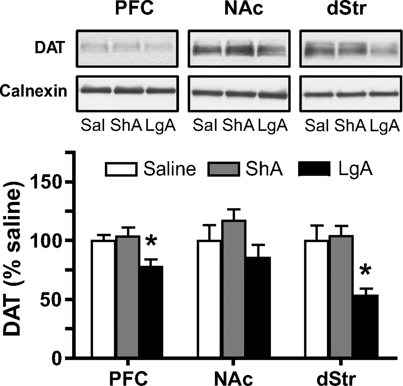
Experiment 3: Extended Meth Self-Administration Reduced the Level of DAT in the PFC and dSTR.
In addition to tissue levels of dopamine and serotonin, the effects of Meth self-administration on the levels of monoamine transporters were investigated in a separate group of animals. For the 2-week Meth maintenance period in experiment 3, animals in the long-access group had a total daily Meth intake of 6.69 ± 0.77 mg/kg (n = 6; mean ± S.E.M. over the last 3 days of self-administration), whereas short access animals had a total daily Meth intake of 1.20 ± 0.12 mg/kg (n = 5). As depicted in Fig. 4, long- but not short-access Meth administration significantly reduced DAT protein levels in both the PFC [F(2,15) = 5.53; p = 0.043] and dSTR [F(2,15) = 5.25; p = 0.021]. There was no change of DAT protein detected in the NAc in any group of rats. In contrast, no changes of total protein levels of norepinephrine or serotonin transporters were detected in surveyed brain areas (Fig. 5; Table 2). Further investigation revealed that Meth did not produce persistent access-dependent changes in a number of protein markers typically associated with Meth-induced neurotoxicity (Fig. 5; Table 2). Thus, there was no change in tyrosine hydroxylase (a dopamine synthetic enzyme), GFAP (a marker of astrocyte activation and proliferation), and Iba-1 (a marker of activated microglia).

DAT protein levels in the PFC, NAc, and dSTR in animals with a history of chronic Meth self-administration and extinction. Representative immunoblots (top insets) depict immunoreactivity of DAT in total cellular homogenates. DAT immunoreactivity was normalized to calnexin and expressed as percentage of the yoked saline control ± S.E.M. (n = 5–6/group). *, p < 0.05 yoked saline control versus long-access Meth. Sal, yoked saline; ShA, short-access Meth; LgA, long-access Meth.

Protein levels of tyrosine hydroxylase, GFAP, Iba-1, norepinephrine transporter (NET), and SERT in the PFC, NAc, and dSTR in animals with a history of chronic Meth self-administration and extinction. Representative immunoblots depicting immunoreactivity of each protein in total cellular homogenates. LgA, long-access Meth; Sal, yoked saline; ShA, short-access Meth; TH, tyrosine hydroxylase.
Protein levels of tyrosine hydroxylase, GFAP, Iba-1, norepinephrine transporter, and SERT in rat forebrain were not altered by long or short access to Meth
All data were normalized to calnexin control and expressed as the percentage of the yoked saline control ± S.E.M. (n = 5 − 6).
Discussion
The key finding of the current study is that extended, but not limited, daily self-administered Meth results in persistent decreases in the levels of DAT protein in the PFC and the dSTR. It is important that these decreases are not likely to be a direct consequence of lasting Meth-induced neurotoxicity, because we found only negligible alterations in dopamine metabolism in the PFC and NAc in short-access animals and no changes in markers specific for dopamine terminals or glial cell activation in either group. The down-regulation of DAT activity may relate to lasting functional consequences of prolonged Meth intake, including motivated drug-seeking behavior, as seen by heightened Meth-primed reinstatement in animals with a history of long-access Meth intake.
Animal models of limited-to-extended daily access to Meth self-administration (as applied in the current study) mimic the course of Meth administration in humans much more closely than studies with experimenter-administered Meth. In support of this, an escalation of Meth intake was observed in rats switched to long access (6 h/day) but not in rats maintained on limited access (1 h/day), which is in agreement with previous studies (Kitamura et al., 2006; Rogers et al., 2008). As a result, we observed robust Meth self-administration (70–110 mg/kg over the 2 weeks) in the long-access rats. This pattern arguably resembles a transition from controlled (limited) to uncontrolled (binge and run) Meth use typical for human addicts (Cho and Melega, 2002). In comparison, typical binge experiments use acute intraperitoneal injections in a single day (ranging from one to four single doses of 1–10 mg/kg) that produce cumulative totals ranging from 4 to 40 mg/kg (Quinton and Yamamoto, 2006; Volz et al., 2007).
Furthermore, self-administration models of Meth addiction (in contrast to noncontingent binge models) can also readily address questions of motivated drug-seeking under relapse conditions. It is significant that chronic Meth use in humans is typically associated with high rates of relapse. As reported previously (Rogers et al., 2008), long-access Meth animals displayed a significant enhancement of Meth-primed reinstatement, a finding similar to that seen after chronic cocaine self-administration (Mantsch et al., 2004; Kippin et al., 2006). In the current study, reinstatement tests with a range of priming doses (0.3–3.0 mg/kg) were performed after 10 days of extinction to assess potential long-term changes in sensitivity to drug effects. Long-access animals readily reinstated to a low priming dose of Meth (0.3 mg/kg) that failed to elicit reinstatement in animals with a history of short access. Although more doses would need to be tested to make a definitive statement about any potential shifts in the dose-response curve, the pattern of behavior in the present study suggests a leftward shift in long-access animals that is indicative of increased sensitivity to a drug prime. As opposed to Meth-primed reinstatement in which differences between short- and long-access animals were evident, there was no difference in cue reactivity between groups. The lack of difference in cue reactivity has been reported previously (Rogers et al., 2008) and may reflect the extensive overtraining with the cues that all animals experienced, regardless of daily access duration. Additional forms of conditioned cued reinstatement (e.g., contextual cues) or changes in cue parameters are warranted in future studies to reveal possible access-dependent differences in relapse.
Both animal and human data have shown previously that chronic Meth results in long-term changes in the function of dopaminergic and serotonergic systems in the brain (for reviews, see Chang et al., 2007; Volz et al., 2007). However, multiple interpretations exist regarding the nature of these changes (compensatory adaptations versus neuronal damage), as well as the degree to which they exhibit recovery over the prolonged drug-free period (Volkow et al., 2001; Wang et al., 2004; McCann et al., 2008). In the current study, dopamine and serotonin levels were assessed after prolonged withdrawal from Meth self-administration. Although animals with a history of extended Meth access display altered motivational and cognitive performance well after cessation of Meth (Rogers et al., 2008; this study), no corresponding changes in dopamine or serotonin levels were detected in the forebrain areas examined. It is interesting that across measures, the only changes suggestive of altered monoamine activity were changes in dopamine metabolism in the ventral PFC and NAc core of rats with short (but not long) access to Meth. Changes in DOPAC or the DOPAC:dopamine ratio have been used as estimates of dopamine transmission, with lower relative production of DOPAC indicating reduced transmission. The significance of the apparent reduction in dopamine transmission in the short-access-only group is not clear. Because activity in the ventral PFC has been strongly linked to extinction learning and relapse (Peters et al., 2008), differences in drug seeking for groups with varied drug access histories may be related to cortical dopaminetransmission.
Although long-access Meth failed to produce lasting changes in monoamine levels, extended Meth resulted in a significant down-regulation of DAT levels in the PFC and dSTR as measured 2 weeks after the end of self-administration. This finding is congruent with clinical reports that have found long-term declines in the levels of DAT in the PFC and dSTR of abstinent Meth users (Sekine et al., 2001, 2003; McCann et al., 2008). Previous studies in animals after Meth self-administration have found only transient changes in DAT and other dopaminergic terminal markers (Stefanski et al., 2002; Shepard et al., 2006). However, these studies only used short-access Meth experience (Stefanski et al., 2002) and shorter total duration of self-administration (Shepard et al., 2006). The current data suggest that more extensive Meth exposure, akin to that seen in chronic human Meth addicts, is necessary for the persistent DAT decrease to occur.
High, neurotoxic doses of Meth (up to 40 mg/kg/daily) delivered in noncontingent binge-type regimens also result in prolonged decreases in striatal DAT levels (Volz et al., 2007). Decreased DAT due to Meth-induced neurotoxicity is typically accompanied by changes in other markers of dopamine terminal damage, such as depletion of striatal dopamine, decrease in tyrosine hydroxylase, activation of glia, and oxidative stress (Thomas et al., 2004; Quinton and Yamamoto, 2006). Given that the maximum daily drug intake of rats with extended access to intravenous Meth was within the range of 6 to 7 mg/kg, DAT decrease was not likely due to Meth-induced dopamine terminal toxicity, particularly because no changes in a number of dopamine terminal markers were detected after chronic Meth self-administration. In some previous studies, repeated administration of below-toxic doses of Meth led to decreased DAT levels in the dSTR but in the absence of significant depletion of striatal dopamine levels (O'Neil et al., 2006; Bjorklund et al., 2008). Therefore, we suggest that decreased DAT levels in the PFC and dSTR represent a neuroadaptation resulting from chronic Meth effects directly on DAT regulation rather than from Meth-induced dopaminergic toxicity. In this regard, it is intriguing that postmortem studies in human Meth addicts have found reduced levels of dopamine terminal markers in the striatum (probably associated with an acute dopamine depletion caused by Meth overdose), but no signs of dopamine terminal degeneration (Wilson et al., 1996; Moszczynska et al., 2004). However, the possibility that the current Meth paradigm did produce some monoaminergic toxicity, which recovered (except for DAT) by the time of tissue analysis, cannot be fully excluded. Therefore, our future studies will evaluate possible signs of Meth-induced cellular toxicity after shorter withdrawal periods (e.g., 24 h).
It is significant that DAT changes occurred in the PFC and dSTR, because these brain structures are part of the circuitry activated during the reinstatement of extinguished cocaine seeking (McFarland and Kalivas, 2001; Fuchs et al., 2006). Therefore, vulnerability to reinstate previously extinguished drug seeking may be associated with decreased DAT levels in the PFC and dSTR as observed in the current study. Decreases in DAT have also been observed in the dSTR of depressed patients displaying a lack of “normal” reward-motivated behavior or anhedonia (Sarchiapone et al., 2006). It is interesting that anhedonia is also one of the symptoms typically associated with withdrawal from chronic Meth use (McGregor et al., 2005). Furthermore, it is important to note that long- but not short-access animals showed a decrease in DAT levels in the PFC and dSTR and that only long-access animals exhibited drug-induced behavioral dysregulations, such as escalation and greater reinstatement of Meth seeking. However, whether and how decreased DAT levels affect extracellular dopamine levels and reinstatement of Meth seeking is not clear. Therefore, future studies will investigate additional changes in cortical and striatal dopamine function (dopamine levels, function of DAT, and vesicular monoamine transporters) in the context of the cognitive and motivational deficits that result from extended Meth self-administration. Alternatively, other factors need to be considered when characterizing neuroadaptations underlying post-Meth behavioral deficits. First, extinction learning itself could play a role in neuroadaptations involved in drug seeking (Sutton et al., 2003). Thus, comparison of animals with a history of extinction versus abstinence after Meth self-administration will be critical. Second, other neurotransmitters not investigated in the current study are probably involved in Meth-induced alterations. In particular, glutamate is involved in Meth-induced toxicity (Quinton and Yamamoto, 2006), Meth reward, and reinforcement (Kim and Jang, 1997; Gass et al., 2009) and reinstatement of Meth seeking (Gass et al., 2009).
Taken together, the present findings suggest that extended Meth self-administration followed by extinction leads to increased Meth-primed reinstatement of drug seeking and decreased DAT in the PFC and dSTR in the absence of persisting changes in dopamine, serotonin, and dopamine metabolism in cortical and striatal subregions. By gaining a greater understanding of these critical substrates in a relevant animal model of Meth addiction, potential targets for therapeutic intervention may be identified and tested.
Acknowledgments
We thank Jason Rogers for technical assistance.
Footnotes
-
This work was supported in part by the Translational Research in Addiction Center at Medical University of South Carolina; the National Institutes of Health National Institute on Drug Abuse [Grant P20-DA022658]; and the National Institutes of Health National Center for Research Resources [Grant CO6-RR015455] (Extramural Research Facilities Program).
-
This work has been presented previously at the 38th Annual Meeting of the Society for Neuroscience, in 2008 Abstract Viewer/Itinerary Planner; 2008 Nov 15–19; Washington, DC. Rocha A, Pacchioni A, and Kalivas PW (2008) Role of the dorsal and ventral prefrontal cortex in cue- and drug-induced reinstatement following extended access to methamphetamine. Program number 358.13; and Schwendt M, Rogers JL, McGinty JF, and See RE (2008) Extended-access to methamphetamine self-administration produces region-specific changes in dopamine transporter signaling assembly in rat forebrain. Program number 358.13.
-
Article, publication date, and citation information can be found at http://jpet.aspetjournals.org.
doi:10.1124/jpet.109.155770
-
ABBREVIATIONS:
-
M.S. and A.R. contributed equally to this work.
- Received May 4, 2009.
- Accepted July 30, 2009.
- © 2009 by The American Society for Pharmacology and Experimental Therapeutics
References
- Armstrong and Noguchi, 2004.↵
- Bjorklund et al., 2008.↵
- Bongiovanni and See, 2008.↵
- Chang et al., 2007.↵
- Cho and Melega, 2002.↵
- Dalley et al., 2007.↵
- Fuchs et al., 2006.↵
- Fumagalli et al., 1998.↵
- Gass et al., 2009.↵
- Haughey et al., 2000.↵
- Kim and Jang, 1997.↵
- Kippin et al., 2006.↵
- Kitamura et al., 2006.↵
- Mantsch et al., 2004.↵
- McCann et al., 2008.↵
- McFarland and Kalivas, 2001.↵
- McGregor et al., 2005.↵
- Moszczynska et al., 2004.↵
- Nordahl et al., 2003.↵
- Numachi et al., 2007.↵
- O'Neil et al., 2006.↵
- Peters et al., 2008.↵
- Prudencio et al., 2002.↵
- Quinton and Yamamoto, 2006.↵
- Rogers et al., 2008.↵
- Sarchiapone et al., 2006.↵
- Scott et al., 2007.↵
- Sekine et al., 2001.↵
- Sekine et al., 2003.↵
- Sekine et al., 2006.↵
- Shaham et al., 2000.↵
- Shepard et al., 2006.↵
- Stefanski et al., 2002.↵
- Sutton et al., 2003.↵
- Szumlinski et al., 2000.↵
- Thomas et al., 2004.↵
- Volkow et al., 2001.↵
- Volz et al., 2007.↵
- Wang et al., 2004.↵
- Wilson et al., 1996.↵


