Article Text

Abstract
OBJECTIVES Different prevalences of generalised osteoarthritis (GOA) in patients with knee and hip OA have been reported. The aim of this investigation was to evaluate radiographic and clinical patterns of disease in a hospital based population of patient subgroups with advanced hip and knee OA and to compare the prevalence of GOA in patients with hip or knee OA, taking potential confounding factors into account.
METHODS 420 patients with hip OA and 389 patients with knee OA scheduled for unilateral total joint replacement in four hospitals underwent radiographic analysis of ipsilateral and contralateral hip or knee joint and both hands in addition to a standardised interview and clinical examination. According to the severity of radiographic changes in the contralateral joints (using Kellgren-Lawrence ⩾ grade 2 as case definition) participants were classified as having either unilateral or bilateral OA. If radiographic changes of two joint groups of the hands (first carpometacarpal joint and proximal/distal interphalangeal joints defined as two separate joint groups) were present, patients were categorised as having GOA.
RESULTS Patients with hip OA were younger (mean age 60.4 years) and less likely to be female (52.4%) than patients with knee OA (66.3 years and 72.5% respectively). Intensity of pain and functional impairment at hospital admission was similar in both groups, while patients with knee OA had a longer symptom duration (median 10 years) compared with patients with hip OA (5 years). In 41.7% of patients with hip OA and 33.4% of patients with knee OA an underlying pathological condition could be observed in the replaced joint, which allowed a classification as secondary OA. Some 82.1% of patients with hip and 87.4% of patients with knee OA had radiographic changes in their contralateral joints (bilateral disease). The prevalence of GOA increased with age and was higher in female patients. GOA was observed more often in patients with knee OA than in patients with hip OA (34.9% versus 19.3%; OR=2.24; 95% CI: 1.56, 3.21). Adjustment for the different age and sex distribution in both patient groups, however, takes away most of the difference (OR=1.32; 95% CI: 0.89, 1.96).
CONCLUSION The crude results confirm previous reports as well as the clinical impression of GOA being more prevalent in patients with advanced knee OA than in patients with advanced hip OA. However, these different patterns might be attributed to a large part to a different distribution of age and sex in these hospital based populations.
- hip osteoarthritis
- knee osteoarthritis
- hand osteoarthritis
- generalised osteoarthritis
Statistics from Altmetric.com
Because of its high prevalence and morbidity, osteoarthritis (OA) is a major cause of impairment and disability among the elderly and therefore a considerable burden both to the individual patient and to society.1 2 Besides commonly affecting the cervical and lumbar spine, most epidemiological studies report a disease predilection for lower extremity weight bearing joints and certain hand joints.3 OA of the knee and of the hip probably have greater social cost and more associated disability than degenerative changes of other joints.4 5
Nevertheless, little is known about the aetiology of the disease, patterns of joint distribution, natural course and predictors of outcome: the identification of involved subjects depends on slowly increasing as well as inconstant signs or symptoms. In addition, the lack of clearly defined disease parameters and a high proportion of asymptomatic patients in early stages makes the design and interpretation of results from non-invasive investigations in community studies more difficult. A reasonable alternative is the investigation of patient subgroups with established disease, where relevant hypotheses can be tested in an appropriate setting. We therefore conducted an epidemiological investigation in the southwestern part of Germany to evaluate radiographic and clinical patterns of advanced hip and knee OA, to assess possible associations with underlying risk factors and to compare predictors of outcome in both disease groups: in a first, cross sectional part of the study, patients scheduled for unilateral hip or knee joint replacement because of severe OA underwent extensive non-invasive clinical investigations, which allow an analysis of the distribution of age, sex, and different clinical as well as radiographic patterns of involvement. In particular, we were interested in the prevalence of generalised osteoarthritis (GOA) in patients with advanced hip and patients with advanced knee OA, as different studies have yielded conflicting results regarding an association between OA of hands, hips, and knees.
Methods
Between January 1995 and December 1996, consecutive patients attending four hospitals (Departments of Orthopaedic Surgery and Traumatology of the University of Ulm; Baumann Orthopaedic Hospital Stuttgart; Hessing Orthopaedic Hospital Augsburg) in the south west of Germany with symptomatic OA of the hip or knee joint severe enough to warrant unilateral total hip or knee arthroplasty were recruited into the study. The following inclusion criteria had to be fulfilled: white race; age under 76 years; absence of malignancies, inflammatory disease or corticosteroid medication; no previous contralateral joint replacement (as the natural course of OA should be investigated in the contralateral, unoperated joint within a consecutive follow up study). The indication for surgical treatment in all patients was painful OA of a hip or knee joint with radiographic changes of at least grade 3 or 4 according to Kellgren and Lawrence.6 The presence of rheumatoid arthritis was excluded by clinical investigation according to ARA criteria.7 The study design was approved by the local ethics committee of the University of Ulm.
CLINICAL INVESTIGATION
After having obtained written informed consent, all patients underwent a detailed investigation to obtain the following baseline data: a standardised interview gathered information on demographic data, duration and severity of symptoms (current hip or knee pain with walking, climbing stairs and/or in resting position indicated on a 100 mm visual analogue scale) as well as drug use. The previous history of the affected joint to be replaced (referred to hereafter as the “ipsilateral joint”) and of the contralateral joint (date and nature of trauma, conservative and surgical treatment of congenital or acquired joint disorders known as secondary causes of OA) was reported in detail.
To measure the degree of functional impairment and to identify the severity of disease in patients with hip and knee OA we included joint specific scores (Lequesne8 and Danielson9score in hip OA, Lequesne8 and Knee-Society Score10 in knee OA) as well as additional algofunctional scores (validated German versions of the “Funktionsfragebogen Hannover (FFbH)”11 and “Western Ontario and McMaster Universities (WOMAC)12 questionnaire”).
RADIOGRAPHIC ASSESSMENT
In patients scheduled for hip joint arthroplasty, a plain radiograph of the pelvis (supine anteroposterior) was obtained and the ipsilateral as well as the contralateral hip joint were examined: overall grading of degenerative changes was performed according to the criteria as described by Kellgren and Lawrence (K and L)6and in an additionally published atlas.13 Separately, for each hip individual radiographic features were scored according to Laneet al 14: lateral and medial joint space narrowing (JSN) as well as lateral and medial osteophytes were graded from 0 to 3 for increasing severity; subchondral acetabular sclerosis, femoral or acetabular cysts and femoral head deformity were noted as absent (grade 0) or present (grade 1).
To identify established secondary patterns of hip OA, all radiographs were assessed for evidence of previous capital epiphyseolysis (identification of a “tilt deformity”15 16) and the presence of a shallow acetabulum with incomplete coverage of the femoral head pointing to underlying hip dysplasia.
In patients undergoing knee replacement anteroposterior weight bearing views in extension and supine lateral views (40 degrees of flexion) of ipsilateral as well as contralateral knee were performed. In addition to K and L overall grading6 17 individual radiographic features of knee OA were determined using the “Baltimore Longitudinal Study of Aging (BLSA)” atlas as described by Scott et al 18: osteophytes and JSN were graded separately for medial and lateral compartments from 0 to 3; medial and lateral tibial subchondral sclerosis, osteophytes of tibial spines and chondrocalcinosis were noted as absent (0) or present (1).
The radiographic reading of all hip and knee films was exclusively performed by a single trained observer (KPG) after documentation of sufficient reproducibility (intra-class correlation coefficients for intraobserver and interobserver reliability of the K and L score of 0.88/0.88 in hip OA and 0.93/0.83 in knee OA) in a reliability study before the present investigation.19 20
In addition to the radiographs of weight bearing joints, we obtained bilateral posteroanterior hand radiographs from most study participants. The first carpometacarpal (CMC) joint as well as all proximal interphalangeal (PIP) and distal interphalangeal (DIP) joints were evaluated regarding JSN, osteophytes and subchondral sclerosis according to an atlas published by Altman et al.21
CLASSIFICATION TOOLS
In hip and knee joints radiographic classification of OA severity was performed with the K and L score,6 13 while hand films were evaluated with a new hand score.
Hip as well as knee osteoarthritis was recorded as present in any joint scored as K and L grade 2 or more. As all ipsilateral hips and knees (operated joints) had to present advanced OA to fulfil inclusion criteria, the definition of unilateral or bilateral OA depended on the severity of degenerative changes in the contralateral joint: in unilateral OA the contralateral changes did not exceed Kellgren grade 1, while in bilateral OA severity grades of ⩾ 2 had to be recorded.
For identification of polyarticular disease subsets the classification criteria of Hart et al 22 were slightly modified: according to their definition of generalised OA (GOA) we looked for radiographic changes in hand films, where PIP joints, DIP joints, and carpometacarpal (CMC) joints were recorded as separate joint regions. But because of the importance of JSN in the definition of hand OA as well as the assessment of progression we scored joints dichotomously as having OA if JSN ⩾ grade 2 alone or alternatively JSN grade 1 together with osteophytes and/or sclerosis ⩾ grade 2 was present. The definition of GOA required involvement of at least two DIP or PIP joints and at least one CMC joint in addition to an osteoarthritic knee or hip joint. One single trained observer (SK) performed the reading again after documentation of sufficient reproduciblity (κ coefficient for intraobserver and interobserver reliability of GOA classification of 0.54, 0.73 respectively) before the investigation.
To distinguish secondary from primary (idiopathic) OA subsets, radiographic findings and data from patient’s past medical history were included: self reported hip and knee joint disorders in the medical history (infection, avascular necrosis and osteochondritis, haemorrhagic diathesis, traumatic events with radiologically and/or surgically confirmed structural joint lesions) as well as radiographic sequelae of slipped femoral capital epiphysis (SCFE) and acetabular dysplasia (DDH) in pelvic radiographs were recorded as evidence of secondary OA.
DATA ANALYSIS
The frequency of GOA was compared between patients with knee OA and patients with hip OA: crude as well as age and sex adjusted prevalences of the different OA patterns were calculated. To adjust for age and sex, we used direct standardisation on the overall number of patients in each stratum. Finally, multivariate logistic regression analyses were used to estimate the odds ratios (OR) and their 95 per cent confidence intervals (CI) for the odds of GOA in patients with knee OA compared with patients with hip OA, adjusting for age (continuous), sex, body mass index (weight in kg/squared height in m, continuous), and the presence of hypertension (systolic blood pressure ⩾ 160 mm Hg or diastolic blood pressure ⩾ 95 mm Hg or use of antihypertensive medication), diabetes (history or use of oral antidiabetics or insulin) and gout (history or use of uricosuric or uricostatic medication).
All statistical analyses were performed using Statistical Analysis Systems (version 6.10), SAS Institute, Cary, North Carolina.
Results
During the two year recruitment period a total of 2153 patients were referred to the participating study centres for unilateral total hip or total knee joint replacement because of advanced OA. According to the inclusion criteria outlined above, 1037 were eligible. The reasons for exclusion were age > 75 years (n=450), previous total joint replacement on the contralateral side (n=330), postponed surgery because of medical or organisational reasons (n=249), inflammatory arthropathies (n=69), and malignancies (n=18). Of the 1037 eligible cases, 809 patients (78.5%) could be recruited for this investigation. As the interview and examination had to be performed on the day of admission for the surgical procedure (scheduled for the next day), recruitment of 212 eligible patients (20.4%) was not possible because of time constraints. Another 16 patients (1.5%) refused to participate. Mean age (63.8 years) and sex distribution (65% female) of these 228 cases were not different from the recruited group.
The study population consisted of 420 patients with hip OA (200 men and 220 women) and 389 patients with knee OA (107 men and 282 women).
Table 1 shows the distribution of demographic and clinical characteristics in both groups of patients. Patients with knee OA (mean age 66.3 years) were on average about six years older than patients with hip OA (60.5 years) and more likely to be women (72.5% women in the knee group compared with 52.4% women in the hip group).
Baseline characteristics of study population (demographics, symptoms, functional impairment, and prevalence of radiographic osteoarthritis)
Joint specific functional rating of hip OA patients with the Danielsson score revealed a mean pain intensity of 80% and a reduction of range of motion and functional capacity by 60% and 40% in the ipsilateral joint. In patients with knee OA, function of the ipsilateral joint was measured with the Knee Society score (mean rating 19.2 points, SD 15.7 points). While overall functional impairment (as measured by Lequesne, WOMAC, and FFbH indices) and severity of symptoms (mean intensity of pain as measured on a VAS) did not show a substantial difference in both groups of patients at the time of hospital referral, patients with knee OA complained of a longer symptom duration in the ipsilateral joint (median 10 years) as compared with participants with hip OA (5 years).
Table 2 shows the identification of underlying conditions as predisposing factors for hip and knee OA: 170 hips (41.7%) and 126 knees (33.4%) could be classified as having secondary OA. While many hips (25.0%) showed sequelae of acetabular dysplasia (DDH), a history of joint trauma was the main predisposing cause for secondary knee OA (28.7%).
Clinically or radiographically identified predisposing conditions for OA of ipsilateral, surgically resected joint in patients with hip and knee OA
A detailed radiographic assessment of severity and distribution of osteoarthritic changes in both hips or knees was possible in 736 patients (91% of study participants), as previous surgery, post-traumatic changes or severe joint deformity did not allow determination of all individual features in some joints. The prevalence of GOA could be assessed in 640 patients (79%), as not all study participants consented to additional hand radiographs. In those 169 patients, who refused hand radiography, the mean age (62.2 years) as well as sex distribution (62.9% women) and frequency of bilateral OA (80.9%) were not significantly different from the findings in all other patients.
Most study participants showed radiographic osteoarthritis of the ipsilateral as well as the contralateral joint: only 72 patients of the hip group and 42 of the knee group (17.9% and 12.6%) had unilateral OA (K and L grade 0 and 1 in the contralateral hip joint). The remaining 330 hip and 292 knee patients with bilateral OA showed changes grade 2 (40.8% and 29.6%), grade 3 (24.9% and 38.9%), and grade 4 (16.4% and 18.9%) in the contralateral joint. Some 49.4% of hip and 81.2% of knee patients with bilateral disease complained of pain in the contralateral joint, while still 25.0% and 47.6% of patients in the unilateral group reported pain without significant contralateral radiographic changes.
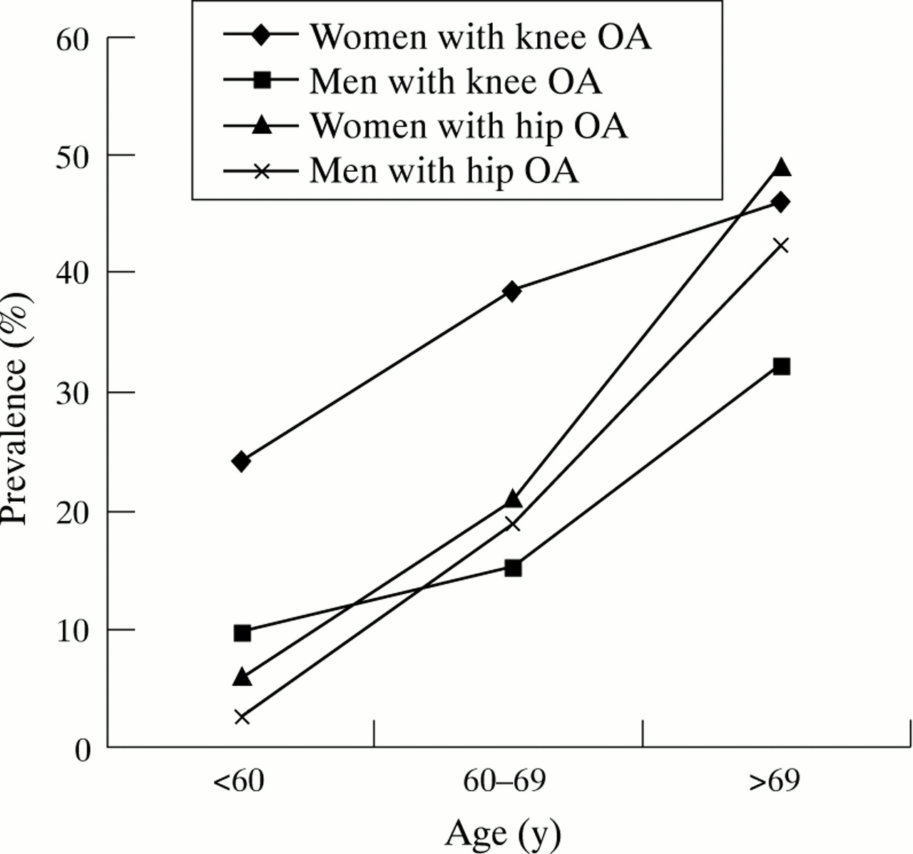
In hands, the DIP and PIP joints were the most frequent sites of radiographic OA involvement in both patient groups (66.4% of patients showed DIP or PIP OA), followed by CMC joints (radiographic changes ⩾ K and L grade 2 in 28.8% of patients). The prevalence of hand OA increased with age in all subgroups and was higher in female patients than in male patients (fig 1).

Age and sex specific prevalence of generalised OA (GOA) in patients with advanced hip and knee OA.
According to our definition of GOA, 26.8% of all patients had polyarticular disease and the prevalence of GOA in the knee group (34.9%) was higher than in the hip group (19.3%). Although distribution of crude data shows a higher frequency of GOA in patients with knee OA compared with hip OA (OR 2.24, CI 1.56, 3.21), this mainly reflects the higher age of knee patients, as no significant independent association persists after adjusting for age and other confounding variables (OR 1.22, CI 0.81, 1.85) in multivariable analyses (table 3). Results remained essentially unchanged after exclusion of patients with secondary OA. As hip and knee patients differed with respect to distribution of age and sex, results regarding laterality, hand involvement and generalisation were standardised for these two influencing factors (fig 2): in contrast with the crude differences (table 1), the age and sex standardised prevalence of GOA was similar in patients with hip and knee OA (23.9% and 28.2%).
Prevalence of GOA (generalised osteoarthritis) in patients with knee OA compared with patients with hip OA

Age and sex standardised comparison of the prevalence of generalised OA, CMC OA, DIP or PIP OA, and bilateral hip or knee OA in patients with advanced hip and knee OA.
Table 4 shows the association of age and sex with GOA in patients with hip and knee OA. A significant association between an age over 65 years and polyarticular disease could be observed in patients with hip as well as knee OA, although the association was stronger in patients with hip disease. In contrast, the association of sex with generalised OA was stronger in patients with knee OA and even more so in a subgroup of patients with primary disease.
Adjusted odds ratios (OR) and 95 % confidence intervals (CI) for the association of age and sex with generalised OA (GOA) derived from multiple logistic regression analyses
Discussion
The main purpose of this investigation was to assess and compare the frequency of GOA in patients with advanced hip or knee OA. We observed a high prevalence of GOA in both subgroups, which is age and sex dependent. After adjusting for these two main confounding factors, the prevalence of generalised OA was similar in patients with hip and knee OA.
Since Kellgren and Moore created the term “generalised osteoarthritis” in 1952,23 the concept of an inherited tendency to multiple joint involvement in OA has been debated extensively. Most authors agree that GOA involves hand joints, the neck, the lower back, and the knees. Initial surveys mainly described patterns of radiographic GOA in population based samples17 24 27 without an analysis of associations between specific joint groups. More recent reports tried to assess type and strength of associations predominantly in patients with knee OA26-33 and their results seem to indicate, that knee OA fits prominently into the concept of GOA.
The inclusion of hip OA into the concept of GOA, however, is more controversial. Since the observation of Kellgren23 34that the hip is rarely involved in GOA, several other investigators also failed to show a high prevalence of multiple joint involvement in patients with hip OA.35-38 On the other hand, Rohet al 39 found a significantly increased frequency of radiographic hand OA in patients with hip OA, compared with normal subjects. Hochberg et al 40 could also demonstrate a significant association of hand OA and bilateral as well as unilateral hip OA in their study group of women over the age of 65 years. In a recent case-control study, Croft et al 41 described a prevalence of Heberden nodes in 25–33% of male patients with hip OA, which supports the results of earlier investigations42-44 also supposing an involvement of the hip in GOA.
There are different reasons that might explain the inconsistency of findings concerning the prevalence of GOA in patients with hip OA and patients with knee OA. They include differences in patient selection, case definition, and classification criteria: in studies reporting an association between knee OA and GOA, participants have most often been recruited from the general population, while investigations of hip OA have often been performed in hospital based patient groups. In community studies a selection bias can be avoided, but in hospital case series a selection of symptomatic OA is evident. Additionally, although knees as well as hip joints are both large, weight bearing, lower limb joints, there are differences in design and function, which are likely to correlate with specific risk factor profiles.45 In our patient group we have seen underlying pathological conditions that probably caused secondary OA in 41.7% of hip and 33.4% of knee patients. This proportion is similar to the distribution in other series at least concerning the hip joint,9 46 47 but a wide range of given ratios of idiopathic compared with secondary OA can be observed.15 16 48-50
Further reasons limiting the comparativeness of identified studies are differences in case definition. OA of different sites has been determined in postmortem studies,37 by radiographic analysis alone,39 40 42 and by combination of clinical and radiographic examination.35 36 41 The sensitivity of either method is different and must be considered in the interpretation of results.
Finally the categorisation of patients as having generalised or localised OA depends on certain classification criteria, which have been used inconsistently for case definition in clinical studies.22-24 51 Different definitions will certainly influence the reported prevalences of GOA, and Doherty et al 45 recently outlined the necessity of clear classification criteria, substantiated by association with putative subset characteristics.
In the Chingford study,22 GOA was defined by the presence of OA in proximal/distal finger joints and CMC joints in addition to knee OA. We have extended this definition to the prevalence of OA in both hand groups in addition to hip OA to allow for valid comparison of hip and knee patients. Although crude prevalence of GOA was higher in patients with knee OA than in patients with hip OA, the frequency of GOA was very similar in both groups after adjustment for age and sex. Previous studies suggesting a higher prevalence of GOA in patients with knee OA than in patients with hip OA have often not adjusted for these major confounders.17 23 24 30
In the interpretation of our results some limitations require careful discussion: because of our inclusion criteria we investigated patients with symptomatic OA requiring total joint replacement. This particular design feature limits generalisability of results to early disease stages. Reported prevalences also do not pertain to the general population but to the study group with advanced radiographic changes in at least one knee or hip joint. But associations might be weaker in early stages and it is difficult to offer a comprehensive study design to a greater number of people with no or minor complaints. Although we cannot compare people with and without OA, our approach facilitates comparison of subgroups of patients with unilateral OA, bilateral OA, and GOA. Our data, however, do not allow an analysis of associations between hip and knee OA directly, as the patients underwent radiographs of either both hips or knees only. Information about involvement of hip and knee joints in the same patient can therefore not be presented, but this does not hinder determination of prevalences of GOA according to the most recent definitions (involvement of two separate joint regions in the hand in addition to OA of at least one large peripheral joint).
Our findings suggest that the reported differences in the medical literature regarding the frequency of GOA in patients with hip and knee OA are at least in part because of an inconsistency of design approaches and different demographic characteristics of study groups. A similar prevalence of GOA in our patient groups with symptomatic hip and knee OA after adjusting for age and sex suggests an influence of systemic aetiological factors in both joints. Additional local risk factors can be identified, but they should not exclude the contribution of generalised susceptibility. For example, the different demographic characteristics of study groups may be explained by different risk factor profiles in hip and knee OA, such as the sex dependent prevalence of childhood hip diseases that may lead to an early onset of degenerative changes.
In conclusion, we examined the association between radiographic OA of hands, hips, and knees in a hospital based population consisting of two patient groups with advanced hip and knee disease. The findings indicate that age and sex are strong determinants of GOA in both groups, and that there is no significant difference in the prevalence of GOA between both groups after adjusting for these and additional confounders.
Acknowledgments
This investigation was supported by the German Ministry of Research and Technology (grant 01 EF 9406). We thank all technicians and secretaries (I Ettler, R Kolb, H v Neubeck, R Schubert-Schmitt, B Mack) for assistance throughout the research project. We are grateful for the cooperation of all colleagues in the participating hospitals, and particularly the patients. We would also like to thank P Dieppe for his ideas and support in developing the radiographic hand score.